Department of Medical Oncology, Amsterdam University Medical Centers, Vrije Universiteit, Cancer Center Amsterdam, Amsterdam, The Netherlands.
Department of Pathology, Amsterdam University Medical Centers, Vrije Universiteit, Cancer Center Amsterdam, Amsterdam, The Netherlands.
J Immunother Cancer. 2021 Mar;9(3). doi: 10.1136/jitc-2020-001962.
We previously reported CpG-B injection at the primary tumor excision site prior to re-excision and sentinel node biopsy to result in immune activation of the sentinel lymph node (SLN), increased melanoma-specific CD8 T cell rates in peripheral blood, and prolonged recurrence-free survival. Here, we assessed recruitment and activation of antigen-presenting cell (APC) subsets in the SLN and at the injection site in relation to T cell infiltration.
Re-excision skin specimens from patients with clinical stage I-II melanoma, collected 7 days after intradermal injection of either saline (n=10) or 8 mg CpG-B (CPG7909, n=12), were examined by immunohistochemistry, quantifying immune subsets in the epidermis, papillary, and reticular dermis. Counts were related to flow cytometric data from matched SLN samples. Additional in vitro cultures and transcriptional analyses on peripheral blood mononuclear cells (PBMCs) were performed to ascertain CpG-induced APC activation and chemokine profiles.
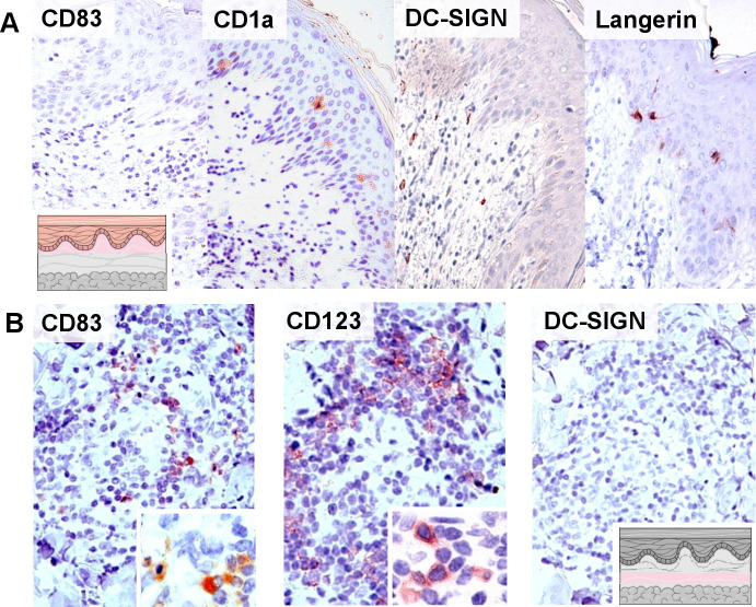
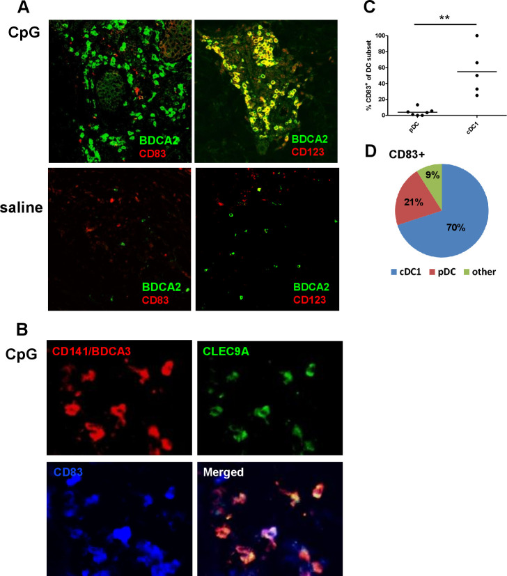
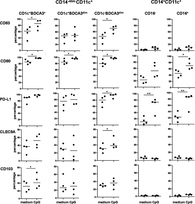
Significant increases in CD83, CD14, CD68, and CD123 APC were observed in the reticular dermis of CpG-B-injected skin samples. Fluorescent double/triple staining revealed recruitment of both CD123BDCA2 plasmacytoid dendritic cells (DCs) and BDCA3/CD141CLEC9A type-1 conventional DC (cDC1), of which only the cDC1 showed considerable levels of CD83 expression. Simultaneous CpG-B-induced increases in T cell infiltration were strongly correlated with both cDC1 and CD14 counts. Moreover, cDC1 and CD14 APC rates in the reticular dermis and matched SLN suspensions were positively correlated. Flow cytometric, transcriptional, and chemokine release analyses of PBMC, on in vitro or in vivo exposure to CpG-B, indicate a role for the activation and recruitment of both cDC1 and CD14 monocyte-derived APCs in the release of CXCL10 and subsequent T cell infiltration.
The CpG-B-induced concerted recruitment of cDC1 and CD14 APC to the injection site and its draining lymph nodes may allow for both the (cross-)priming of T cells and their subsequent homing to effector sites.
我们之前报道过在再次切除和前哨淋巴结活检前,在原发性肿瘤切除部位注射 CpG-B,这导致前哨淋巴结(SLN)的免疫激活,外周血中黑色素瘤特异性 CD8 T 细胞比例增加,并延长无复发生存期。在这里,我们评估了与 T 细胞浸润相关的 SLN 和注射部位抗原呈递细胞(APC)亚群的募集和激活。
从临床 I-II 期黑色素瘤患者的再次切除皮肤标本中,在皮内注射生理盐水(n=10)或 8mg CpG-B(CPG7909,n=12)后 7 天收集,通过免疫组织化学进行检查,定量表皮、乳头层和网状真皮中的免疫亚群。计数与匹配的 SLN 样本的流式细胞术数据相关。还对外周血单核细胞(PBMC)进行了额外的体外培养和转录分析,以确定 CpG 诱导的 APC 激活和趋化因子谱。
在 CpG-B 注射皮肤样本的网状真皮中,观察到 CD83、CD14、CD68 和 CD123 APC 的显著增加。荧光双重/三重染色显示,BDCA2 浆细胞样树突状细胞(pDC)和 BDCA3/CD141CLEC9A 1 型传统树突状细胞(cDC1)的募集,其中只有 cDC1 显示出相当水平的 CD83 表达。同时,CpG-B 诱导的 T 细胞浸润增加与 cDC1 和 CD14 计数呈强相关性。此外,网状真皮和匹配的 SLN 悬浮液中的 cDC1 和 CD14 APC 率呈正相关。对 PBMC 的体外或体内暴露于 CpG-B 进行流式细胞术、转录和趋化因子释放分析表明,cDC1 和 CD14 单核细胞来源的 APC 的激活和募集在 CXCL10 的释放和随后的 T 细胞浸润中起作用。
CpG-B 诱导的 cDC1 和 CD14 APC 到注射部位及其引流淋巴结的协同募集可能允许 T 细胞的(交叉)启动及其随后向效应部位的归巢。