From the Sorbonne University (E.P., M.T., F.-X.L., V.A.G.R., M.B.d.l.M., C.B., G.B., B.B., B.S.), Paris Brain Institute; Imaging Department (E.P.), Foundation A. de Rothschild Hospital, Paris; Paris-Saclay University (M.T., B.K., M.B.), CEA, Orsay; and Assistance Publique des Hôpitaux de Paris (B.B., B.S.), France.
Neurology. 2021 Apr 6;96(14):e1865-e1875. doi: 10.1212/WNL.0000000000011700. Epub 2021 Mar 18.
To explore in vivo innate immune cell activation as a function of the distance from ventricular CSF in patients with multiple sclerosis (MS) using [F]-DPA714 PET and to investigate its relationship with periventricular microstructural damage, evaluated by magnetization transfer ratio (MTR), and with trajectories of disability worsening.
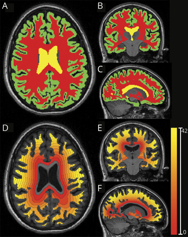
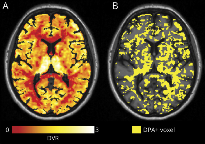
Thirty-seven patients with MS and 19 healthy controls underwent MRI and [F]-DPA714 TSPO dynamic PET, from which individual maps of voxels characterized by innate immune cell activation (DPA+) were generated. White matter (WM) was divided in 3-mm-thick concentric rings radiating from the ventricular surface toward the cortex, and the percentage of DPA+ voxels and mean MTR were extracted from each ring. Two-year trajectories of disability worsening were collected to identify patients with and without recent disability worsening.
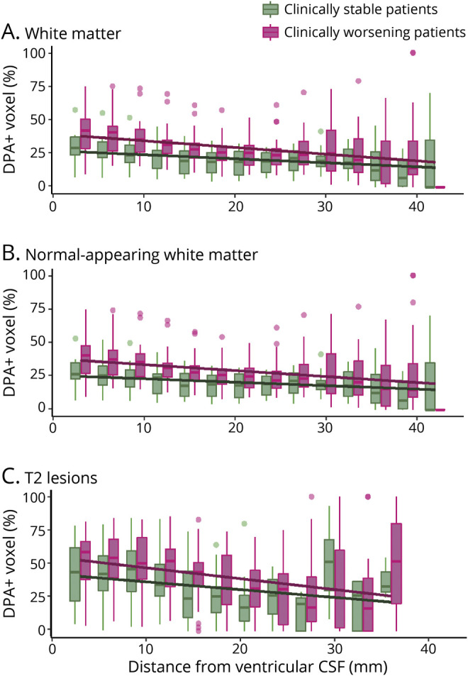
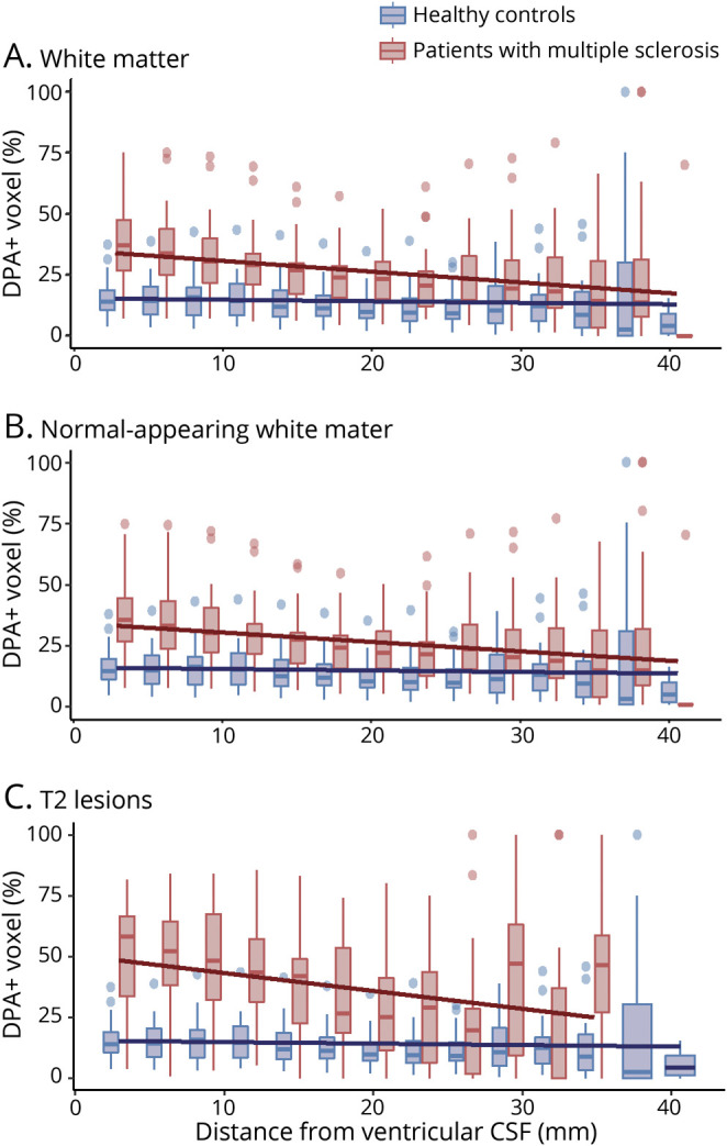
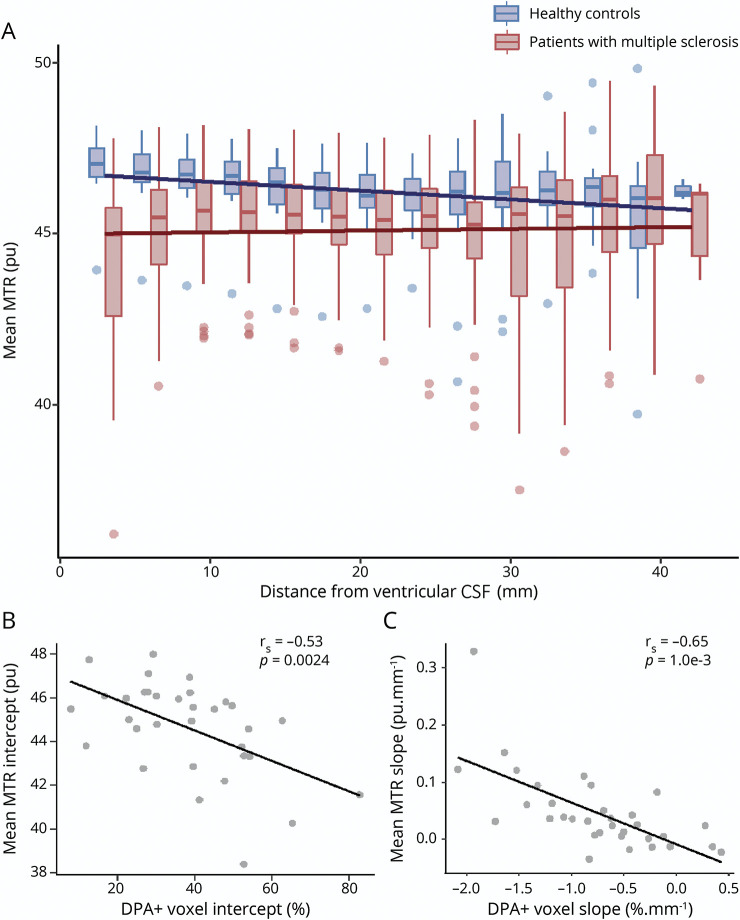
The percentage of DPA+ voxels was higher in patients compared to controls in the periventricular WM ( = 6.10e-6) and declined with increasing distance from ventricular surface, with a steeper gradient in patients compared to controls ( = 0.001). This gradient was found in both periventricular lesions and normal-appearing WM. In the total WM, it correlated with a gradient of microstructural tissue damage measured by MTR ( = -0.65, = 1.0e-3). Compared to clinically stable patients, patients with disability worsening were characterized by a higher percentage of DPA+ voxels in the periventricular normal-appearing WM ( = 0.025).
Our results demonstrate that in MS the innate immune cell activation predominates in periventricular regions and is associated with microstructural damage and disability worsening. This could result from the diffusion of proinflammatory CSF-derived factors into surrounding tissues.
利用 [F]-DPA714 PET 探索多发性硬化症(MS)患者脑室 CSF 距离不同时体内固有免疫细胞激活情况,并研究其与脑室周围微观结构损伤(通过磁化传递率(MTR)评估)和残疾恶化轨迹的关系。
37 例 MS 患者和 19 名健康对照者接受 MRI 和 [F]-DPA714 TSPO 动态 PET 检查,从这些检查中生成固有免疫细胞激活(DPA+)的体素个体图谱。将白质(WM)分为从脑室表面向皮质放射的 3mm 厚的同心环,并从每个环中提取 DPA+体素的百分比和平均 MTR。收集了 2 年的残疾恶化轨迹,以确定有无近期残疾恶化的患者。
与对照组相比,患者的脑室周围 WM 中 DPA+体素的百分比更高( = 6.10e-6),且随着与脑室表面距离的增加而降低,患者的梯度比对照组更陡峭( = 0.001)。该梯度在脑室周围病变和正常表现 WM 中均存在。在整个 WM 中,它与 MTR 测量的微观结构组织损伤梯度相关( = -0.65, = 1.0e-3)。与临床稳定的患者相比,残疾恶化的患者脑室周围正常表现 WM 中的 DPA+体素百分比更高( = 0.025)。
我们的研究结果表明,在 MS 中固有免疫细胞的激活主要发生在脑室周围区域,与微观结构损伤和残疾恶化有关。这可能是由于促炎 CSF 衍生因子扩散到周围组织所致。