Doheny Eye Centers-UCLA, Pasadena, CA, USA.
Department of Ophthalmology and Visual Sciences, University of Maryland School of Medicine, Baltimore, MD, USA.
Sci Rep. 2021 Mar 18;11(1):6344. doi: 10.1038/s41598-021-85010-1.
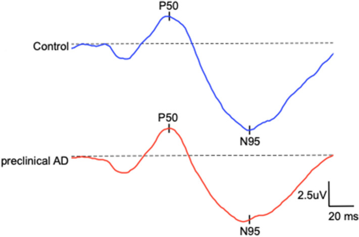
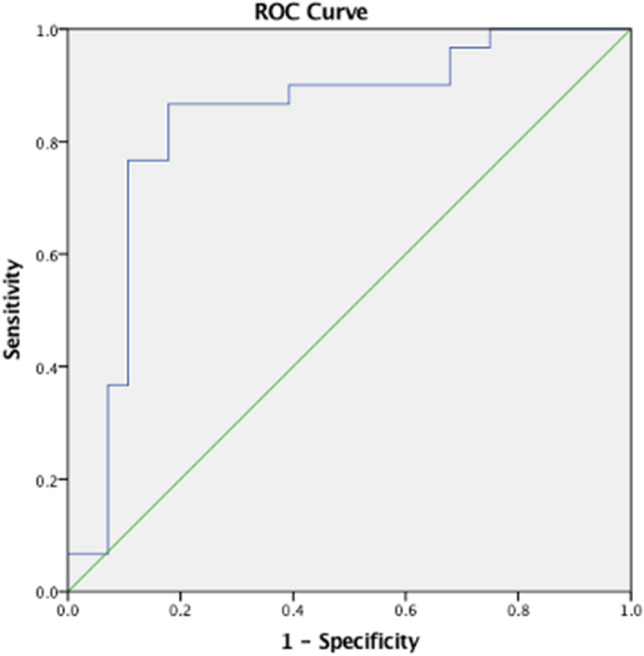
The current study evaluated retinal function using electroretinography (ERG) in cognitively healthy (CH) participants with preclinical Alzheimer's disease (AD), as classified by cerebral spinal fluid (CSF) Aβ/Tau ratio. Individuals with normal retinal morphology ascertained by spectral-domain optical coherence tomography were enrolled. Full-field ERG, pattern PERG, and photopic negative response (PhNR) were performed in 29 adult participants (58 eyes). Amplitude and implicit times of the ERG wave components were analyzed. Preclinical AD participants showed marked retinal ganglion cell dysfunction relative to controls. The PhNR was significantly diminished in preclinical AD relative to controls. PhNR amplitude and N95 implicit time differentiated CH individuals with CSF biomarkers of AD pathology with 87% sensitivity and 82% specificity. These quantitative electrophysiologic findings expand our understanding of early retinal functional changes that precede cognitive decline in AD. Retinal ganglion cell dysfunction, as detected by ERG, may be a clinically useful, non-invasive in vivo biomarker for early disease detection, which is necessary for ultimately pursuing early intervention.
本研究通过检测认知健康(CH)伴有临床前阿尔茨海默病(AD)个体的视网膜功能,使用视网膜电图(ERG)评估脑脊液(CSF)中 Aβ/Tau 比值。通过频域光相干断层扫描(OCT)确认个体的视网膜形态正常后,对 29 名成年参与者(58 只眼)进行全视野 ERG、图形 PERG 和明适应负向反应(PhNR)检测。分析 ERG 波成分的振幅和潜伏期。与对照组相比,临床前 AD 参与者的视网膜神经节细胞功能明显受损。与对照组相比,临床前 AD 患者的 PhNR 明显降低。PhNR 振幅和 N95 潜伏期可区分 CSF 生物标志物提示 AD 病理的 CH 个体,其敏感性为 87%,特异性为 82%。这些定量电生理发现扩展了我们对 AD 认知能力下降前早期视网膜功能变化的理解。ERG 检测到的视网膜神经节细胞功能障碍可能是一种有用的、非侵入性的体内生物标志物,可用于早期疾病检测,这对于最终追求早期干预是必要的。