Doheny Eye Institute, Los Angeles, California, United States.
Department of Ophthalmology, David Geffen School of Medicine at the University of California Los Angeles, Los Angeles, California, United States.
Invest Ophthalmol Vis Sci. 2019 Apr 1;60(5):1491-1500. doi: 10.1167/iovs.18-25966.
To provide a histopathologic, morphometric analysis of the retina in Alzheimer's disease (AD).
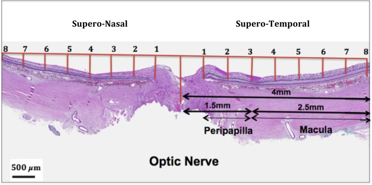
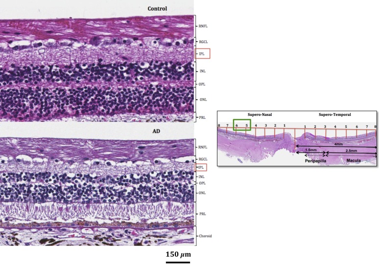
Human postmortem retinas from eight patients with AD (mean age: 80 ± 12.7 years) and from 11 age-matched controls (mean age: 78 ± 16.57 years) were analyzed. The retinas were sampled from the superior quadrant on both the temporal and nasal sides with respect to the optic nerve. Thickness of the inner and outer layers involving the retinal nerve fiber layer (RNFL), retinal ganglion cell layer (RGCL), inner plexiform layer (IPL), inner nuclear layer (INL), and outer nuclear layer (ONL) were measured and compared between controls and AD. A total of 16 measurements of retinal thickness were acquired for each layer.
RNFL thinning supero-temporally was significant closest to the optic nerve (∼35% thickness reduction; P < 0.001). Supero-nasally, RNFL was thinner throughout all points (∼40% reduction; P < 0.001). Supero-temporally, RGCL thinning was pronounced toward the macula (∼35% thickness reduction; P < 0.001). Supero-nasally, RGCL showed uniform thinning throughout (∼35% reduction; P < 0.001). IPL thinning supero-temporally was statistically significant in the macula (∼15% reduction; P < 0.01). Supero-nasal IPL featured uniform thinning throughout (∼25% reduction; P < 0.001). Supero-temporally, INL and ONL thinning were pronounced toward the macula (∼25% reduction; P < 0.01). Supero-nasally, INL and ONL were thinner throughout (∼25% reduction; P < 0.01).
Our study revealed marked thinning in both the inner and outer layers of the retina. These quantified histopathologic findings provide a more comprehensive understanding of the retina in AD than previously reported.
对阿尔茨海默病(AD)患者的视网膜进行组织病理学、形态计量学分析。
对 8 例 AD 患者(平均年龄:80 ± 12.7 岁)和 11 名年龄匹配的对照者(平均年龄:78 ± 16.57 岁)的死后视网膜进行分析。视网膜取自视神经颞侧和鼻侧上方的象限。测量并比较对照组和 AD 组中视网膜神经纤维层(RNFL)、视网膜神经节细胞层(RGCL)、内丛状层(IPL)、内核层(INL)和外核层(ONL)的内层和外层厚度。对各层的视网膜厚度进行了 16 次测量。
最靠近视神经的上方颞侧的 RNFL 变薄显著(厚度减少约 35%;P < 0.001)。上方鼻侧的 RNFL 各部位均变薄(厚度减少约 40%;P < 0.001)。上方颞侧的 RGCL 向黄斑区变薄明显(厚度减少约 35%;P < 0.001)。上方鼻侧的 RGCL 整体变薄(厚度减少约 35%;P < 0.001)。上方颞侧 IPL 在黄斑区变薄有统计学意义(厚度减少约 15%;P < 0.01)。上方鼻侧 IPL 整体变薄(厚度减少约 25%;P < 0.001)。上方颞侧的 INL 和 ONL 向黄斑区变薄明显(厚度减少约 25%;P < 0.01)。上方鼻侧的 INL 和 ONL 整体变薄(厚度减少约 25%;P < 0.01)。
本研究揭示了视网膜内层和外层的明显变薄。这些量化的组织病理学发现比以前报道的更全面地了解 AD 患者的视网膜。