Hart Nadav J, Koronyo Yosef, Black Keith L, Koronyo-Hamaoui Maya
Department of Neurosurgery, Maxine Dunitz Neurosurgical Research Institute, Cedars-Sinai Medical Center, 127 S. San Vicente Blvd., Los Angeles, 90048, CA, USA.
Department of Biomedical Sciences, Cedars-Sinai Medical Center, 110 George Burns Rd., Los Angeles, CA, 90048, USA.
Acta Neuropathol. 2016 Dec;132(6):767-787. doi: 10.1007/s00401-016-1613-6. Epub 2016 Sep 19.
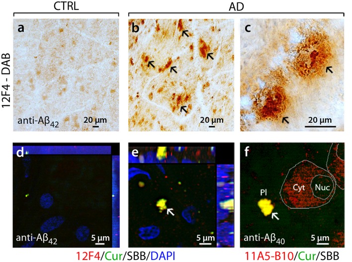
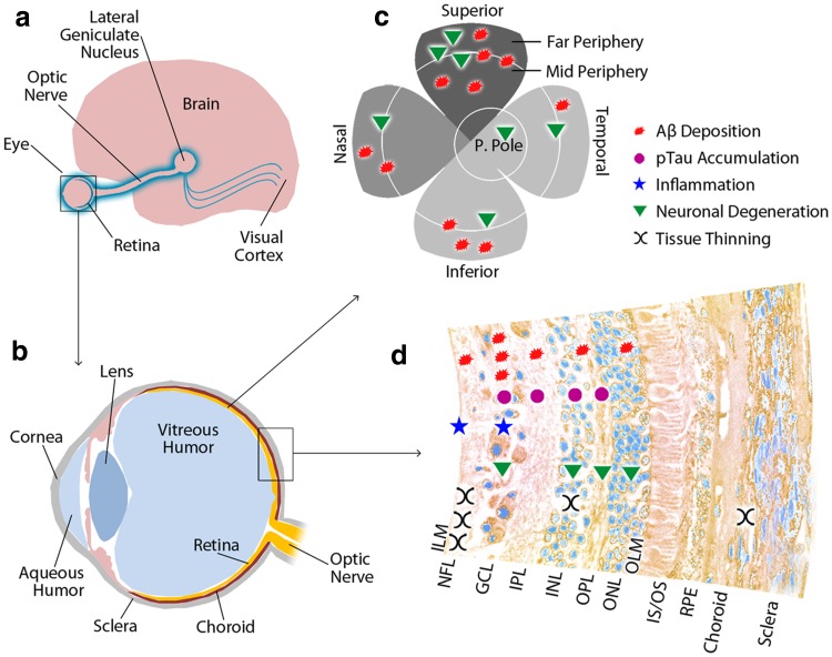
Although historically perceived as a disorder confined to the brain, our understanding of Alzheimer's disease (AD) has expanded to include extra-cerebral manifestation, with mounting evidence of abnormalities in the eye. Among ocular tissues, the retina, a developmental outgrowth of the brain, is marked by an array of pathologies in patients suffering from AD, including nerve fiber layer thinning, degeneration of retinal ganglion cells, and changes to vascular parameters. While the hallmark pathological signs of AD, amyloid β-protein (Aβ) plaques and neurofibrillary tangles (NFT) comprising hyperphosphorylated tau (pTau) protein, have long been described in the brain, identification of these characteristic biomarkers in the retina has only recently been reported. In particular, Aβ deposits were discovered in post-mortem retinas of advanced and early stage cases of AD, in stark contrast to non-AD controls. Subsequent studies have reported elevated Aβ peptides, morphologically diverse Aβ plaques, and pTau in the retina. In line with the above findings, animal model studies have reported retinal Aβ deposits and tauopathy, often correlated with local inflammation, retinal ganglion cell degeneration, and functional deficits. This review highlights the converging evidence that AD manifests in the eye, especially in the retina, which can be imaged directly and non-invasively. Visual dysfunction in AD patients, traditionally attributed to well-documented cerebral pathology, can now be reexamined as a direct outcome of retinal abnormalities. As we continue to study the disease in the brain, the emerging field of ocular AD warrants further investigation of how the retina may faithfully reflect the neurological disease. Indeed, detection of retinal AD pathology, particularly the early presenting amyloid biomarkers, using advanced high-resolution imaging techniques may allow large-scale screening and monitoring of at-risk populations.
尽管从历史上看,阿尔茨海默病(AD)被认为是一种局限于大脑的疾病,但我们对它的理解已经扩展到包括脑外表现,越来越多的证据表明眼睛也存在异常。在眼部组织中,视网膜是大脑发育的延伸部分,患有AD的患者会出现一系列病理变化,包括神经纤维层变薄、视网膜神经节细胞变性以及血管参数改变。虽然AD的标志性病理特征——由高度磷酸化的tau(pTau)蛋白组成的淀粉样β蛋白(Aβ)斑块和神经原纤维缠结(NFT)——早已在大脑中被描述,但直到最近才报道在视网膜中发现了这些特征性生物标志物。特别是,在AD晚期和早期病例的死后视网膜中发现了Aβ沉积物,这与非AD对照组形成了鲜明对比。随后的研究报告了视网膜中Aβ肽升高、形态多样的Aβ斑块以及pTau。与上述发现一致,动物模型研究报告了视网膜Aβ沉积物和tau病变,通常与局部炎症、视网膜神经节细胞变性和功能缺陷相关。这篇综述强调了越来越多的证据表明AD在眼睛中表现出来,尤其是在视网膜中,而视网膜可以直接进行非侵入性成像。AD患者的视觉功能障碍传统上归因于有充分记录的脑部病变,现在可以重新审视为视网膜异常的直接结果。随着我们继续在大脑中研究这种疾病,眼部AD这一新兴领域值得进一步研究视网膜如何忠实地反映这种神经疾病。事实上,使用先进的高分辨率成像技术检测视网膜AD病理,特别是早期出现的淀粉样生物标志物,可能有助于对高危人群进行大规模筛查和监测。