Schurz Natascha, Sariaslani Lydia, Altmann Patrick, Leutmezer Fritz, Mitsch Christoph, Pemp Berthold, Rommer Paulus, Zrzavy Tobias, Berger Thomas, Bsteh Gabriel
Department of Neurology, Medical University of Vienna, Vienna, Austria.
Department of Ophthalmology, Medical University of Vienna, Vienna, Austria.
Eye Brain. 2021 Mar 12;13:59-69. doi: 10.2147/EB.S295610. eCollection 2021.
Retinal layer thickness parameters measured by optical coherence tomography (OCT) are emerging biomarkers of neuroaxonal degeneration and inflammation in multiple sclerosis (MS). We aimed to evaluate the value of retinal layer thickness for prediction of disability worsening and relapse in a real-world MS cohort.
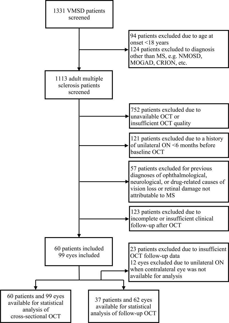
For this longitudinal observational study, we included MS patients with spectral-domain OCT scans available and ≥1 year of clinical follow-up. The value of peripapillary retinal nerve fiber layer (pRNFL), macular ganglion-cell-and-inner-plexiform-layer (GCIPL) and inner nuclear layer (INL) thickness for prediction of disability worsening and relapse during the observation period was tested by multivariate models.
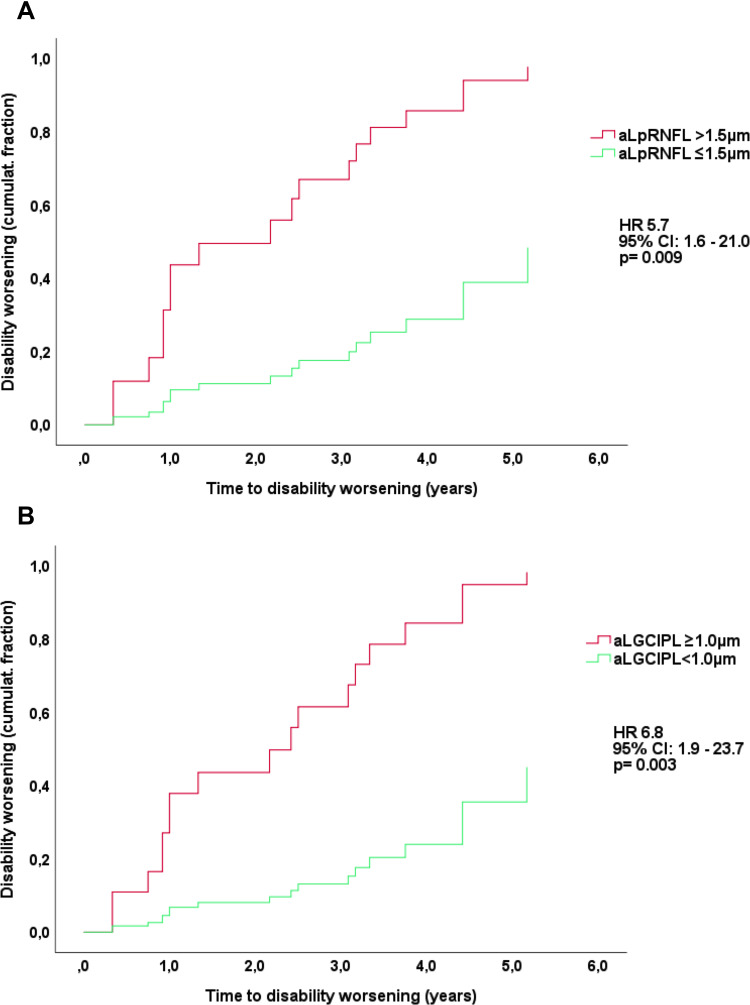
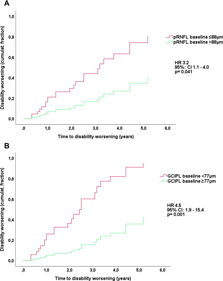
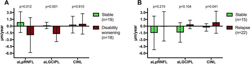
We analyzed 60 MS patients during a mean observation period of 2.9 years (SD 1.8). Lower baseline thickness of GCIPL (cut-off <77µm; HR 4.1, p=0.001) and pRNFL (cut-off ≤88µm; HR 3.1, p=0.019) were associated with an increased risk of disability worsening. Longitudinally, mean thinning rates were -0.8µm/year (SD 1.6) for GCIPL, -0.6µm/year (SD 3.5) for pRNFL. GCIPL thinning ≥1.0µm/year and pRNFL >1.5µm/year is associated with higher likelihood of disability worsening (HR 5.7, p=0.009 and HR 6.8, p=0.003, respectively). INL thickened in patients with relapse by a mean 0.9µm while thinning by 0.3µm in patients without relapse (p=0.04). In multivariate analyses, INL thickening was associated with an increased probability of relapse (OR 17.8, p=0.023).
Cross-sectional and longitudinal measurement of GCIPL and pRNFL thinning is reliable as a biomarker of disability worsening in a real-world setting. Change of INL thickness is a promising marker of relapse, i.e. inflammatory activity.
光学相干断层扫描(OCT)测量的视网膜层厚度参数正成为多发性硬化症(MS)神经轴突退变和炎症的生物标志物。我们旨在评估在真实世界的MS队列中,视网膜层厚度对预测残疾恶化和复发的价值。
在这项纵向观察性研究中,我们纳入了有光谱域OCT扫描且临床随访≥1年的MS患者。通过多变量模型测试视乳头周围视网膜神经纤维层(pRNFL)、黄斑神经节细胞及内丛状层(GCIPL)和内核层(INL)厚度对观察期内残疾恶化和复发的预测价值。
我们在平均2.9年(标准差1.8)的观察期内分析了60例MS患者。GCIPL基线厚度较低(临界值<77μm;风险比4.1,p=0.001)和pRNFL(临界值≤88μm;风险比3.1,p=0.019)与残疾恶化风险增加相关。纵向来看,GCIPL的平均变薄率为-0.8μm/年(标准差1.6),pRNFL为-0.6μm/年(标准差3.5)。GCIPL变薄≥1.0μm/年和pRNFL>1.5μm/年与残疾恶化的可能性更高相关(风险比分别为5.7,p=0.009和风险比6.8,p=0.003)。复发患者的INL平均增厚0.9μm,未复发患者的INL平均变薄0.3μm(p=0.04)。在多变量分析中,INL增厚与复发概率增加相关(比值比17.8,p=0.023)。
在真实世界中,GCIPL和pRNFL变薄的横断面和纵向测量作为残疾恶化的生物标志物是可靠的。INL厚度变化是复发即炎症活动的一个有前景的标志物。