• 文献检索
  • 文档翻译
  • 深度研究
  • 学术资讯
  • Suppr Zotero 插件Zotero 插件
  • 邀请有礼
  • 套餐&价格
  • 历史记录
应用&插件
Suppr Zotero 插件Zotero 插件浏览器插件Mac 客户端Windows 客户端微信小程序
定价
高级版会员购买积分包购买API积分包
服务
文献检索文档翻译深度研究API 文档MCP 服务
关于我们
关于 Suppr公司介绍联系我们用户协议隐私条款
关注我们

Suppr 超能文献

核心技术专利:CN118964589B侵权必究
粤ICP备2023148730 号-1Suppr @ 2026

文献检索

告别复杂PubMed语法,用中文像聊天一样搜索,搜遍4000万医学文献。AI智能推荐,让科研检索更轻松。

立即免费搜索

文件翻译

保留排版,准确专业,支持PDF/Word/PPT等文件格式,支持 12+语言互译。

免费翻译文档

深度研究

AI帮你快速写综述,25分钟生成高质量综述,智能提取关键信息,辅助科研写作。

立即免费体验

视网膜层变薄预示着复发型多发性硬化症的治疗失败。

Retinal layer thinning predicts treatment failure in relapsing multiple sclerosis.

机构信息

Department of Neurology, Medical University of Vienna, Vienna, Austria.

Department of Neurology, Medical University of Innsbruck, Innsbruck, Austria.

出版信息

Eur J Neurol. 2021 Jun;28(6):2037-2045. doi: 10.1111/ene.14829. Epub 2021 Apr 2.

DOI:10.1111/ene.14829
PMID:33735479
原文链接:https://pmc.ncbi.nlm.nih.gov/articles/PMC8251588/
Abstract

BACKGROUND AND PURPOSE

Peripapillary retinal nerve fiber layer (pRNFL) and macular ganglion cell plus inner plexiform layer (GCIPL) thinning are markers of neuroaxonal degeneration in multiple sclerosis (MS), which is reduced by disease-modifying treatment (DMT). We aimed to investigate the potential of pRNFL and GCIPL thinning for prediction of DMT failure in relapsing MS (RMS).

METHODS

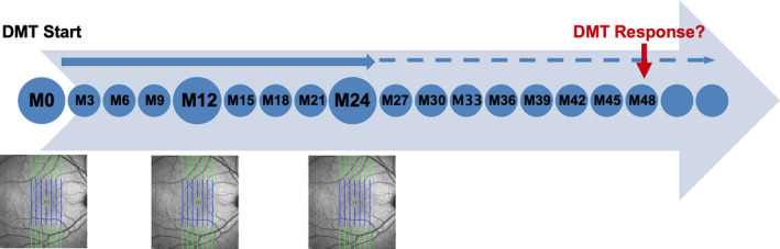
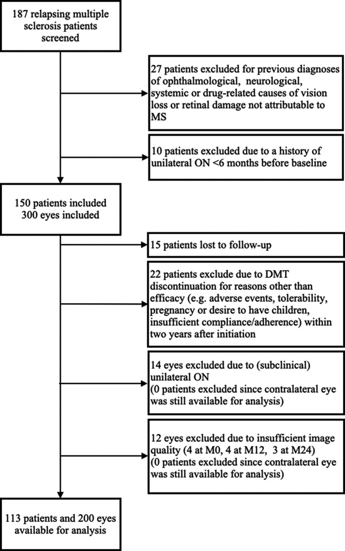
In this 4-year prospective observational study on 113 RMS patients, pRNFL and GCIPL were measured at DMT initiation and after 12 months (M12) and 24 months (M24). Treatment failure was defined as 6-month confirmed Expanded Disability Status Scale (EDSS) progression and/or Symbol Digit Modalities Test (SDMT) worsening. Optimal cutoff values for predicting treatment failure were determined by receiver operating characteristic analyses and hazard ratios (HRs) by multivariable Cox regression adjusting for age, sex, disease duration, EDSS/SDMT, and DMT class.

RESULTS

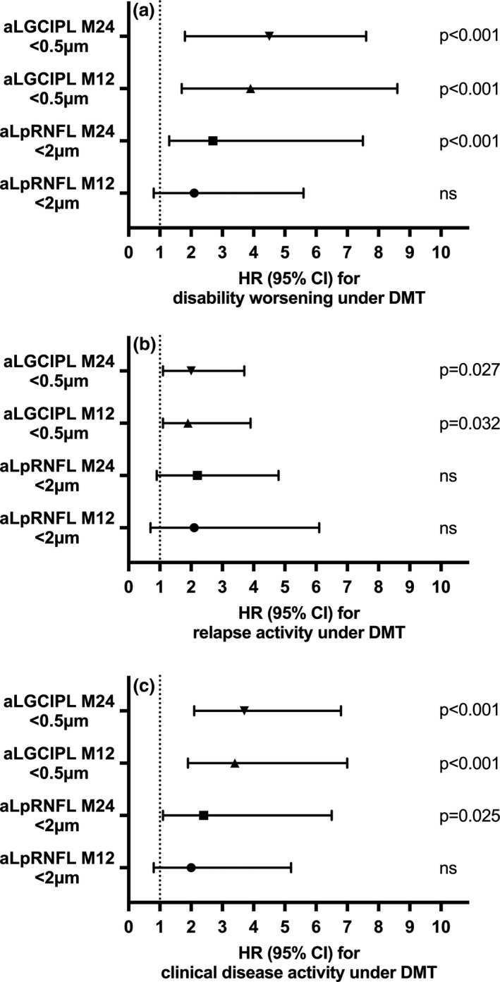
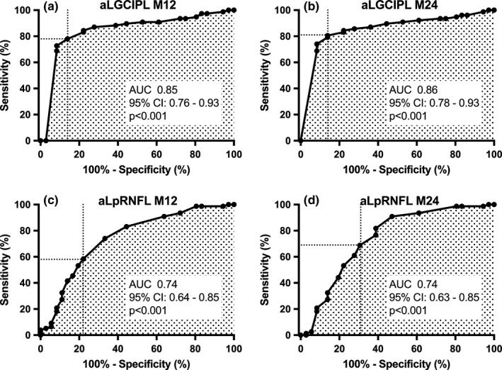
Thinning of GCIPL >0.5 μm/year at M24 showed superior value for treatment failure prediction (HR: 4.5, 95% confidence interval [CI]: 1.8-7.6, p < 0.001; specificity 91%, sensitivity 81%), followed by GCIPL >0.5 μm at M12 (odds ratio [OR]: 3.9, 95% CI: 1.4-6.9, p < 0.001; specificity 85%, sensitivity 78%), and pRNFL ≥2 μm/year at M24 (OR: 3.7, 95% CI: 1.1-6.5, p = 0.023; specificity 84%, sensitivity 69%), whereas pRNFL at M12 was not predictive.

CONCLUSIONS

GCIPL, and to a lesser degree pRNFL, thinning predicts disability progression after DMT initiation and may be a useful and accessible biomarker of treatment failure in RMS.

摘要

背景与目的

视盘周围视网膜神经纤维层(pRNFL)和黄斑神经节细胞加内丛状层(GCIPL)变薄是多发性硬化症(MS)神经轴突变性的标志物,这种变性可被疾病修正治疗(DMT)所减轻。我们旨在研究 pRNFL 和 GCIPL 变薄对预测复发型多发性硬化症(RMS)DMT 失败的潜在价值。

方法

在这项针对 113 例 RMS 患者的 4 年前瞻性观察性研究中,在 DMT 起始时以及 12 个月(M12)和 24 个月(M24)时测量了 pRNFL 和 GCIPL。将 6 个月确认的扩展残疾状况量表(EDSS)进展和/或符号数字模态测试(SDMT)恶化定义为治疗失败。通过接受者操作特征分析确定预测治疗失败的最佳截断值,并通过多变量 Cox 回归调整年龄、性别、疾病持续时间、EDSS/SDMT 和 DMT 类别来确定危险比(HR)。

结果

M24 时 GCIPL 变薄 >0.5μm/年对治疗失败预测具有更高的价值(HR:4.5,95%置信区间[CI]:1.8-7.6,p<0.001;特异性 91%,敏感性 81%),其次是 M12 时 GCIPL >0.5μm(优势比[OR]:3.9,95%CI:1.4-6.9,p<0.001;特异性 85%,敏感性 78%)和 M24 时 pRNFL ≥2μm/年(OR:3.7,95%CI:1.1-6.5,p=0.023;特异性 84%,敏感性 69%),而 M12 时的 pRNFL 则没有预测价值。

结论

GCIPL 和 pRNFL 的变薄程度较小,可预测 DMT 起始后残疾进展,可能是 RMS 中治疗失败的有用且可获得的生物标志物。

https://cdn.ncbi.nlm.nih.gov/pmc/blobs/c413/8251588/18ccd870bb43/ENE-28-2037-g002.jpg
https://cdn.ncbi.nlm.nih.gov/pmc/blobs/c413/8251588/c9ea9f797c9a/ENE-28-2037-g005.jpg
https://cdn.ncbi.nlm.nih.gov/pmc/blobs/c413/8251588/77375251a8d8/ENE-28-2037-g003.jpg
https://cdn.ncbi.nlm.nih.gov/pmc/blobs/c413/8251588/430b8ca58714/ENE-28-2037-g004.jpg
https://cdn.ncbi.nlm.nih.gov/pmc/blobs/c413/8251588/18ccd870bb43/ENE-28-2037-g002.jpg

相似文献

1
Retinal layer thinning predicts treatment failure in relapsing multiple sclerosis.视网膜层变薄预示着复发型多发性硬化症的治疗失败。
Eur J Neurol. 2021 Jun;28(6):2037-2045. doi: 10.1111/ene.14829. Epub 2021 Apr 2.
2
Retinal layer thickness predicts disability accumulation in early relapsing multiple sclerosis.视网膜层厚度可预测早期复发型多发性硬化症患者残疾程度的累积。
Eur J Neurol. 2023 Apr;30(4):1025-1034. doi: 10.1111/ene.15718. Epub 2023 Feb 16.
3
Retinal layer thinning is reflecting disability progression independent of relapse activity in multiple sclerosis.视网膜层变薄反映了多发性硬化症中与复发活动无关的残疾进展。
Mult Scler J Exp Transl Clin. 2020 Oct 29;6(4):2055217320966344. doi: 10.1177/2055217320966344. eCollection 2020 Oct-Dec.
4
Retinal layer thinning as a biomarker of long-term disability progression in multiple sclerosis.视网膜层变薄作为多发性硬化症长期残疾进展的生物标志物。
Mult Scler. 2022 Oct;28(12):1871-1880. doi: 10.1177/13524585221097566. Epub 2022 Jun 2.
5
Normative Data and Conversion Equation for Spectral-Domain Optical Coherence Tomography in an International Healthy Control Cohort.国际健康对照队列中光谱域光学相干断层扫描的参考数据和转换公式。
J Neuroophthalmol. 2022 Dec 1;42(4):442-453. doi: 10.1097/WNO.0000000000001717. Epub 2022 Oct 18.
6
Association of Serum Neurofilament Light Chain With Inner Retinal Layer Thinning in Multiple Sclerosis.血清神经丝轻链与多发性硬化症内视网膜层变薄的关联。
Neurology. 2022 Aug 16;99(7):e688-e697. doi: 10.1212/WNL.0000000000200778. Epub 2022 May 26.
7
Comparative diagnostic accuracy of ganglion cell-inner plexiform and retinal nerve fiber layer thickness measures by Cirrus and Spectralis optical coherence tomography in relapsing-remitting multiple sclerosis.在复发缓解型多发性硬化症中,使用Cirrus和Spectralis光学相干断层扫描测量神经节细胞-内网状层和视网膜神经纤维层厚度的比较诊断准确性。
Biomed Res Int. 2014;2014:128517. doi: 10.1155/2014/128517. Epub 2014 Sep 18.
8
Evaluation of Retinal Layer Thickness Parameters as Biomarkers in a Real-World Multiple Sclerosis Cohort.在真实世界多发性硬化症队列中评估视网膜层厚度参数作为生物标志物
Eye Brain. 2021 Mar 12;13:59-69. doi: 10.2147/EB.S295610. eCollection 2021.
9
Retinal layer thinning for monitoring disease-modifying treatment in relapsing multiple sclerosis-Evidence for applying a rebaselining concept.视网膜层变薄用于监测复发型多发性硬化症的疾病修正治疗——应用重新设定基线概念的证据。
Mult Scler. 2024 Aug;30(9):1128-1138. doi: 10.1177/13524585241267257. Epub 2024 Aug 7.
10
Optical coherence tomography versus other biomarkers: Associations with physical and cognitive disability in multiple sclerosis.光学相干断层扫描与其他生物标志物:与多发性硬化症的身体和认知残疾的关联。
Mult Scler. 2023 Nov;29(13):1540-1550. doi: 10.1177/13524585231198760. Epub 2023 Sep 29.

引用本文的文献

1
The Relationship Between OCT and VEP Parameters with Disability and Disease Duration in Relapsing-Remitting Multiple Sclerosis.复发缓解型多发性硬化症中光学相干断层扫描(OCT)和视觉诱发电位(VEP)参数与残疾及病程的关系
Diagnostics (Basel). 2025 Aug 28;15(17):2181. doi: 10.3390/diagnostics15172181.
2
Retinal Optical Coherence Tomography Longitudinal Measures as Prognostic Biomarkers in Multiple Sclerosis: Systematic Review and Meta-Analysis.视网膜光学相干断层扫描纵向测量作为多发性硬化症的预后生物标志物:系统评价和荟萃分析
Neurol Neuroimmunol Neuroinflamm. 2025 Jul;12(4):e200416. doi: 10.1212/NXI.0000000000200416. Epub 2025 May 27.
3

本文引用的文献

1
Interferon-β Is Less Effective Than Other Drugs in Controlling the Rate of Retinal Ganglion Cell Loss in MS.干扰素-β 在控制 MS 中视网膜神经节细胞损失率方面的效果不如其他药物。
Neurol Neuroimmunol Neuroinflamm. 2021 Feb 17;8(3). doi: 10.1212/NXI.0000000000000971. Print 2021 May.
2
Validation of inter-eye difference thresholds in optical coherence tomography for identification of optic neuritis in multiple sclerosis.光学相干断层扫描中双眼差异阈值用于识别多发性硬化症视神经炎的验证
Mult Scler Relat Disord. 2020 Oct;45:102403. doi: 10.1016/j.msard.2020.102403. Epub 2020 Jul 17.
3
Macular ganglion cell-inner plexiform layer thinning as a biomarker of disability progression in relapsing multiple sclerosis.
Retinal thinning differentiates treatment effects in relapsing multiple sclerosis below the clinical threshold.
视网膜变薄可区分复发型多发性硬化症低于临床阈值的治疗效果。
Ann Clin Transl Neurol. 2025 Feb;12(2):345-354. doi: 10.1002/acn3.52279. Epub 2024 Dec 16.
4
Evolution of retinal degeneration and prediction of disease activity in relapsing and progressive multiple sclerosis.视网膜变性的演变与复发进展性多发性硬化症疾病活动的预测。
Nat Commun. 2024 Jun 19;15(1):5243. doi: 10.1038/s41467-024-49309-7.
5
Effects of Ibudilast on Retinal Atrophy in Progressive Multiple Sclerosis Subtypes: Post Hoc Analyses of the SPRINT-MS Trial.依达拉奉对进展型多发性硬化亚型中视网膜萎缩的影响:SPRINT-MS 试验的事后分析。
Neurology. 2023 Sep 5;101(10):e1014-e1024. doi: 10.1212/WNL.0000000000207551. Epub 2023 Jul 17.
6
Emerging imaging and liquid biomarkers in multiple sclerosis.多发性硬化症中的新兴影像和液体生物标志物。
Eur J Immunol. 2023 Aug;53(8):e2250228. doi: 10.1002/eji.202250228. Epub 2023 May 28.
7
Swept-Source Optical Coherence Tomography Thresholds in Differentiating Clinical Outcomes in a Real-World Cohort of Treatment-Naïve Multiple Sclerosis Patients.在未经治疗的多发性硬化症患者真实世界队列中,扫频源光学相干断层扫描在区分临床结局方面的阈值
Brain Sci. 2023 Mar 31;13(4):591. doi: 10.3390/brainsci13040591.
8
Optical coherence tomography (OCT) measurements and cognitive performance in multiple sclerosis: a systematic review and meta-analysis.光学相干断层扫描(OCT)测量与多发性硬化症的认知表现:一项系统评价和荟萃分析
J Neurol. 2023 Mar;270(3):1266-1285. doi: 10.1007/s00415-022-11449-5. Epub 2022 Nov 17.
9
Optical coherence tomography assessment of axonal and neuronal damage of the retina in patients with familial and sporadic multiple sclerosis.光学相干断层扫描评估家族性和散发性多发性硬化症患者视网膜的轴突和神经元损伤。
Front Neurol. 2022 Sep 16;13:953188. doi: 10.3389/fneur.2022.953188. eCollection 2022.
黄斑神经节细胞-内丛状层变薄可作为复发型多发性硬化残疾进展的生物标志物。
Mult Scler. 2021 Apr;27(5):684-694. doi: 10.1177/1352458520935724. Epub 2020 Jul 2.
4
To treat or not to treat: Sequential individualized treatment evaluation in relapsing multiple sclerosis.治疗还是不治疗:复发型多发性硬化症的序贯个体化治疗评估
Mult Scler Relat Disord. 2020 Apr;39:101908. doi: 10.1016/j.msard.2019.101908. Epub 2019 Dec 23.
5
Association of brain volume loss and long-term disability outcomes in patients with multiple sclerosis treated with teriflunomide.特立氟胺治疗多发性硬化症患者的脑容量损失与长期残疾结局的相关性。
Mult Scler. 2020 Sep;26(10):1207-1216. doi: 10.1177/1352458519855722. Epub 2019 Jun 14.
6
Current therapeutic landscape in multiple sclerosis: an evolving treatment paradigm.多发性硬化症的当前治疗格局:不断发展的治疗范例。
Curr Opin Neurol. 2019 Jun;32(3):365-377. doi: 10.1097/WCO.0000000000000700.
7
Optimal intereye difference thresholds by optical coherence tomography in multiple sclerosis: An international study.光学相干断层扫描在多发性硬化症中的最佳眼间差异阈值:一项国际研究。
Ann Neurol. 2019 May;85(5):618-629. doi: 10.1002/ana.25462. Epub 2019 Apr 10.
8
Peripapillary retinal nerve fibre layer thinning rate as a biomarker discriminating stable and progressing relapsing-remitting multiple sclerosis.视盘周围视网膜神经纤维层变薄率作为鉴别稳定和进展复发缓解型多发性硬化症的生物标志物。
Eur J Neurol. 2019 Jun;26(6):865-871. doi: 10.1111/ene.13897. Epub 2019 Feb 19.
9
'No evidence of disease activity' - is it an appropriate surrogate in multiple sclerosis?“无疾病活动证据”——在多发性硬化症中是否是一个合适的替代指标?
Eur J Neurol. 2018 Sep;25(9):1107-e101. doi: 10.1111/ene.13669. Epub 2018 May 28.
10
Disease-modifying treatments for early and advanced multiple sclerosis: a new treatment paradigm.早期和晚期多发性硬化症的疾病修正治疗:一种新的治疗模式。
Curr Opin Neurol. 2018 Jun;31(3):233-243. doi: 10.1097/WCO.0000000000000561.