Ramjiani Vipul, Mudhar Hardeep-Singh, Julian Thomas, Auger Graham
Department of Ophthalmology, Sheffield Teaching Hospitals NHS Foundation Trust, Royal Hallamshire Hospital, Glossop Rd, England, S10 2JF, Sheffield, UK.
National Specialist Ophthalmic Pathology Service (NSOPS), Dept of Histopathology-E-Floor, Royal Hallamshire Hospital, Glossop Rd, England, S10 2JF, Sheffield, UK.
BMC Ophthalmol. 2021 Mar 19;21(1):138. doi: 10.1186/s12886-021-01895-6.
To report sampling of the trabecular meshwork using the TrabEx+ (MicroSurgical Technology, Redmond, Washington, USA) device in ab interno trabeculectomy. Specifically, this series focusses upon preservation of the trabecular meshwork architecture for assessment of glaucomatous features using common histopathological techniques.
This series features six glaucomatous eyes undergoing TrabEx+ with or without cataract surgery. Three patients had primary open angle glaucoma and the remaining had pigment dispersion glaucoma, ocular hypertension or uveitic glaucoma. Four eyes had simultaneous cataract surgery.
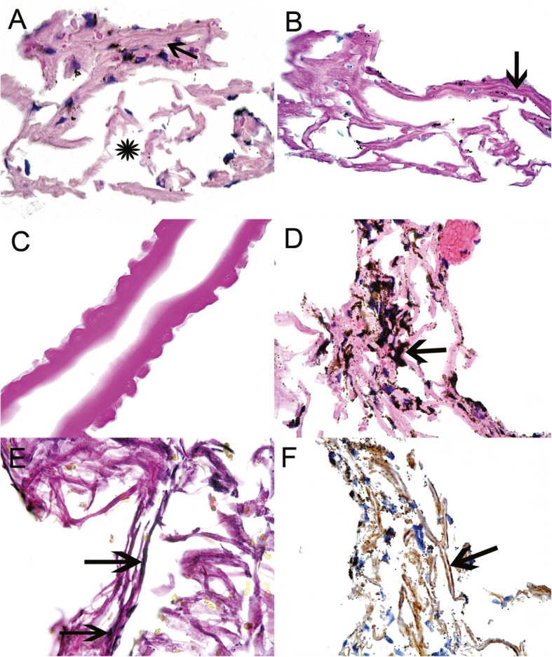
Trabecular meshwork was excised using the TrabEx+ device and retrieved using vitreoretinal forceps. The samples were then processed into formalin-fixed paraffin-embedded 4 micron tissue segments and stained with haematoxylin and eosin, periodic acid-Schiff and elastin Van Gieson. Collagen IV was labelled using immunohistochemistry for the purpose of identifying the basement membrane of trabecular beams.
Presence of trabecular meshwork was confirmed in five of the six samples taken. One of six samples consisted of blood only, but this was expected following early termination of the procedure due to patient restlessness. In the five positive cases trabecular beams with associated trabecular meshwork cells were identified on hematoxylin-eosin and periodic acid-Schiff staining. The beams retained their lamellar structure. The basement membrane underlying the trabecular cells was evident in three specimens, whilst two specimens were of insufficient size for collagen IV labelling.
This case series illustrates that TrabEx+ can be utilised to successfully retrieve trabecular meshwork samples with sufficient architectural perseveration of the tissue to enable histopathological and laboratory analysis.
报告在非穿透性小梁切除术中使用TrabEx+(美国华盛顿州雷德蒙德市显微外科技术公司)设备对小梁网进行取材。具体而言,本系列研究聚焦于保留小梁网结构,以便使用常见的组织病理学技术评估青光眼特征。
本系列研究包括6只接受TrabEx+手术的青光眼患眼,部分患者同时接受或未接受白内障手术。3例患者患有原发性开角型青光眼,其余患者患有色素性青光眼、高眼压症或葡萄膜炎性青光眼。4只眼同时接受了白内障手术。
使用TrabEx+设备切除小梁网,并用玻璃体视网膜镊取出样本。然后将样本处理成福尔马林固定石蜡包埋的4微米组织切片,并用苏木精和伊红、过碘酸希夫和弹性蛋白范吉森染色。为了识别小梁束的基底膜,使用免疫组织化学对IV型胶原进行标记。
在采集的6个样本中,有5个样本证实存在小梁网。6个样本中有1个仅为血液,但这是由于患者躁动导致手术提前终止所预期的结果。在5例阳性病例中,苏木精-伊红和过碘酸希夫染色显示有小梁束及相关小梁网细胞。小梁束保留了其板层结构。3个标本中可见小梁细胞下方的基底膜,而2个标本因尺寸不足无法进行IV型胶原标记。
本病例系列表明,TrabEx+可用于成功获取小梁网样本,该组织具有足够的结构保留,能够进行组织病理学和实验室分析。