Celularity Inc, Florham Park, New Jersey, USA.
Celularity Inc, Florham Park, New Jersey, USA
J Immunother Cancer. 2021 Mar;9(3). doi: 10.1136/jitc-2020-001975.
Tumors often develop resistance to surveillance by endogenous immune cells, which include natural killer (NK) cells. Ex vivo activated and/or expanded NK cells demonstrate cytotoxicity against various tumor cells and are promising therapeutics for adoptive cancer immunotherapy. Genetic modification can further enhance NK effector cell activity or activation sensitization. Here, we evaluated the effect of the genetic deletion of ubiquitin ligase Casitas B-lineage lymphoma pro-oncogene-b (), a negative regulator of lymphocyte activity, on placental CD34 cell-derived NK (PNK) cell cytotoxicity against tumor cells.
Using CRISPR/Cas9 technology, was knocked out in placenta-derived CD34 hematopoietic stem cells, followed by differentiation into PNK cells. Cell expansion, phenotype and cytotoxicity against tumor cells were characterized in vitro. The antitumor efficacy of knockout (KO) PNK cells was tested in an acute myeloid leukemia (HL-60) tumor model in NOD- IL2R gamma (NSG) mice. PNK cell persistence, biodistribution, proliferation, phenotype and antitumor activity were evaluated.
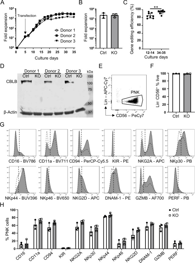
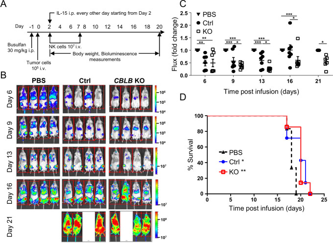
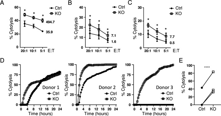
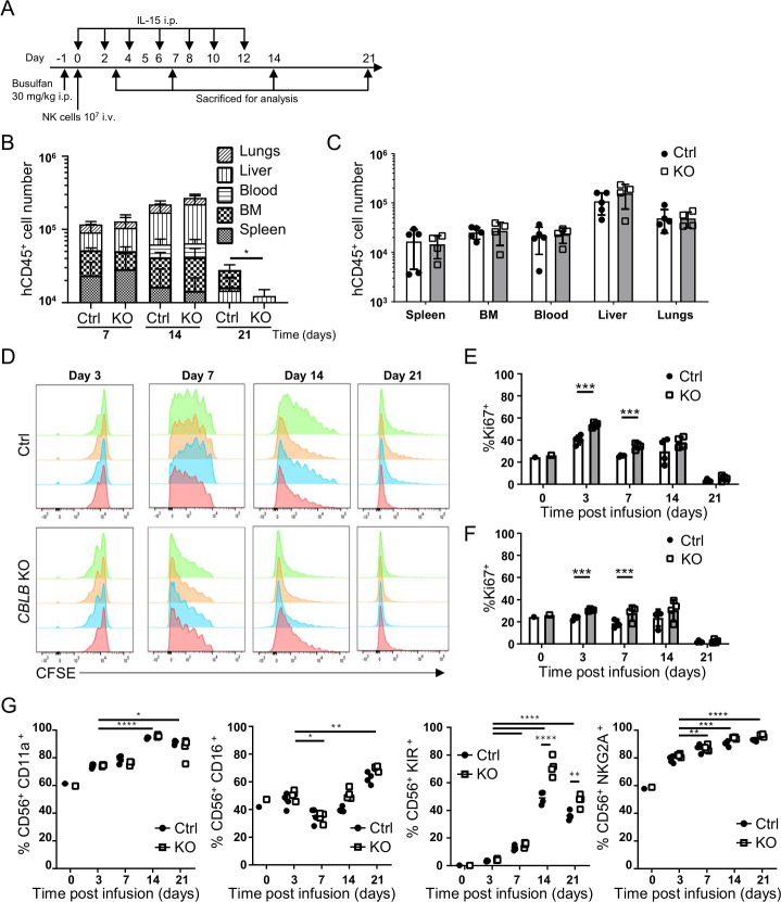
94% of KO efficacy was achieved using CRISPR/Cas9 gene editing technology. KO placental CD34 cells differentiated into PNK cells with high cell yield and >90% purity determined by CD56 CD3 cell identity. Ablation of did not impact cell proliferation, NK cell differentiation or phenotypical characteristics of PNK cells. When compared with the unmodified PNK control, KO PNK cells exhibited higher cytotoxicity against a range of liquid and solid tumor cell lines in vitro. On infusion into busulfan-conditioned NSG mice, KO PNK cells showed in vivo proliferation and maturation as evidenced by increased expression of CD16, killer Ig-like receptors and NKG2A over 3 weeks. Additionally, KO PNK cells showed greater antitumor activity in a disseminated HL60-luciferase mouse model compared with unmodified PNK cells.
ablation increased PNK cell effector function and proliferative capacity compared with non-modified PNK cells. These data suggest that targeting may offer therapeutic advantages via enhancing antitumor activities of NK cell therapies.
肿瘤经常会对包括自然杀伤 (NK) 细胞在内的内源性免疫细胞的监测产生抗性。体外激活和/或扩增的 NK 细胞对各种肿瘤细胞具有细胞毒性,是过继性癌症免疫治疗的有前途的治疗方法。遗传修饰可以进一步增强 NK 效应细胞的活性或激活敏感性。在这里,我们评估了泛素连接酶 Casitas B 细胞淋巴瘤原癌基因-b () 的遗传缺失对胎盘 CD34 细胞衍生的 NK (PNK) 细胞对肿瘤细胞的细胞毒性的影响, Casitas B 细胞淋巴瘤原癌基因-b 是淋巴细胞活性的负调节剂。
使用 CRISPR/Cas9 技术敲除胎盘衍生的 CD34 造血干细胞中的 Casitas B 细胞淋巴瘤原癌基因-b,然后分化为 PNK 细胞。在体外对细胞扩增、表型和对肿瘤细胞的细胞毒性进行了表征。在 NOD-IL2Rγ(NSG)小鼠的急性髓系白血病(HL-60)肿瘤模型中测试了 基因敲除 (KO) PNK 细胞的抗肿瘤功效。评估了 PNK 细胞的持久性、体内分布、增殖、表型和抗肿瘤活性。
使用 CRISPR/Cas9 基因编辑技术,达到了 94%的 KO 效果。CRISPR/Cas9 基因编辑技术敲除 Casitas B 细胞淋巴瘤原癌基因-b 的胎盘 CD34 细胞分化为 PNK 细胞,细胞产量高,纯度>90%,通过 CD56 CD3 细胞鉴定。Casitas B 细胞淋巴瘤原癌基因-b 的缺失不影响细胞增殖、NK 细胞分化或 PNK 细胞的表型特征。与未经修饰的 PNK 对照相比,KO PNK 细胞在体外对一系列液体和固体肿瘤细胞系表现出更高的细胞毒性。在白消安预处理的 NSG 小鼠输注后,KO PNK 细胞在体内增殖和成熟,3 周后 CD16、杀伤免疫球蛋白样受体和 NKG2A 的表达增加。此外,与未经修饰的 PNK 细胞相比,KO PNK 细胞在播散性 HL60-荧光素酶小鼠模型中表现出更强的抗肿瘤活性。
与未经修饰的 PNK 细胞相比,KO 增加了 PNK 细胞的效应功能和增殖能力。这些数据表明,通过增强 NK 细胞疗法的抗肿瘤活性,靶向 Casitas B 细胞淋巴瘤原癌基因-b 可能具有治疗优势。