Su Linglin, Wang Rui, Qiu Tianxin, Wang Jinli, Meng Jinwu, Zhu Jinyue, Wang Deyun, Wu Yi, Liu Jiaguo
MOE Joint International Research Laboratory of Animal Health and Food Safety and Institute of Traditional Chinese Veterinary Medicine, College of Veterinary Medicine, Nanjing Agricultural University, Nanjing 210095, P R China.
MOE Joint International Research Laboratory of Animal Health and Food Safety and Institute of Traditional Chinese Veterinary Medicine, College of Veterinary Medicine, Nanjing Agricultural University, Nanjing 210095, P R China.
Poult Sci. 2021 May;100(5):101032. doi: 10.1016/j.psj.2021.101032. Epub 2021 Feb 9.
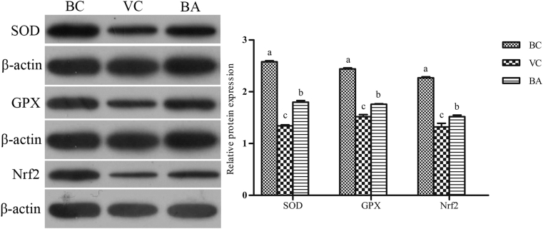
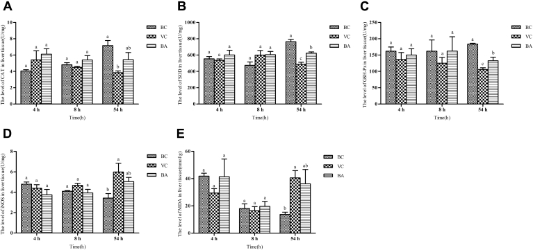
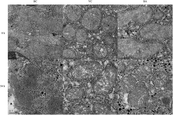
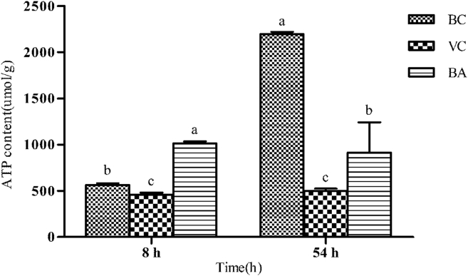
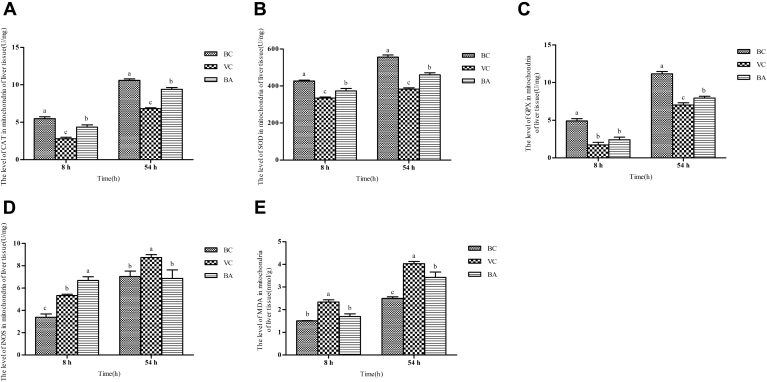
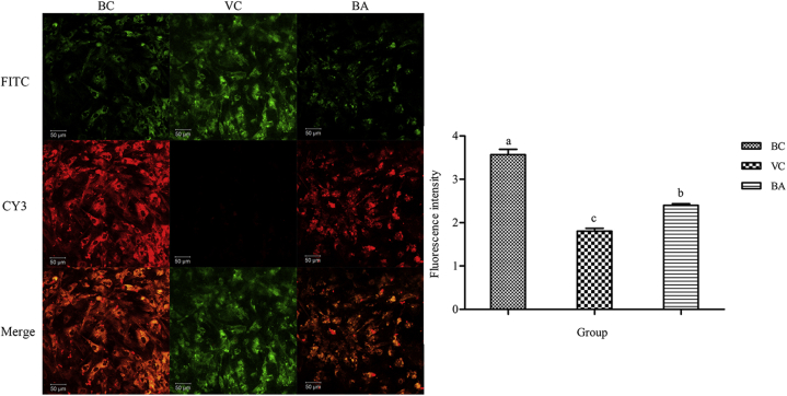
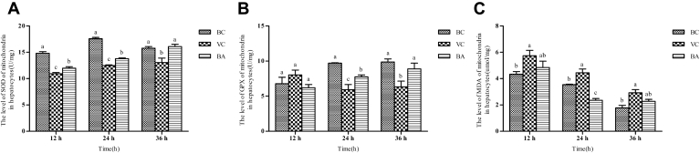
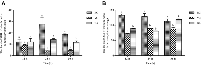
Duck hepatitis A virus type 1 (DHAV-1) is the main pathogen of duck viral hepatitis, but the efficacy of the licensed commercial vaccine needs to be further improved. Therapeutic measures of specific drugs for DHAV-1-infected ducklings need to be urgently developed. Baicalin possesses good antiviral effects. This study aims to investigate the mechanism of baicalin in protecting hepatic mitochondrial function from DHAV-1. The ELISA method was used to detect changes of hepatic and mitochondrial catalase (CAT), superoxide dismutase (SOD), glutathione peroxidase (GPX), inducible nitric oxide synthase (iNOS), adenosine triphosphate (ATP), and malondialdehyde (MDA) levels in vivo and vitro. Hematoxylin and eosin sections and transmission electron microscopy were used to observe liver pathological changes and mitochondrial structural changes. The changes in mitochondrial membrane potential were detected by JC-1 staining method. Western blot and quantitative real-time PCR were employed to analyze the gene and protein expressions in the nuclear erythroid 2-related factor 2 (Nrf2)/antioxidant responsive element (ARE) pathway in duck embryonic hepatocytes infected with DHAV-1. Results showed the administration of baicalin increased the survival rate of ducklings, and alleviated hepatic damage caused by DHAV-1 by enhancing the antioxidant enzyme activities of the liver and mitochondria, including SOD, GPX, CAT, and reducing lipid peroxidative damage (MDA content) and iNOS activities. The mitochondrial ultrastructure changed and the significant increase of ATP content showed that baicalin maintained the structural integrity and ameliorated mitochondrial dysfunction after DHAV-1 infection. In vitro, DHAV-1 infection led to loss of mitochondrial membrane potential and lipid peroxidation and decreased antioxidative enzyme activities (SOD, GPX) and mitochondrial respiratory chain complex activities (succinate dehydrogenase, cytochrome c oxidase). Baicalin relieved the above changes caused by DHAV-1 and activated the gene and protein expressions of Nrf2, which activated ARE-dependent genes including heme oxygenase-1 (HO-1), nicotinamide adenine dinucleotide phosphate quinone oxidoreductase 1 (NQO1), SOD-1, and GPX-1. In addition, baicalin increased the protein expressions of antioxidative enzymes (SOD, GPX). Hence, baicalin protects the liver against oxidative stress in hepatic mitochondria caused by DHAV-1 via activating the Nrf2/ARE signaling pathway.
鸭甲型肝炎病毒1型(DHAV-1)是鸭病毒性肝炎的主要病原体,但现有市售疫苗的效力仍需进一步提高。急需开发针对感染DHAV-1雏鸭的特异性药物治疗措施。黄芩苷具有良好的抗病毒作用。本研究旨在探讨黄芩苷保护肝脏线粒体功能免受DHAV-1侵害的机制。采用ELISA法检测体内外肝脏及线粒体过氧化氢酶(CAT)、超氧化物歧化酶(SOD)、谷胱甘肽过氧化物酶(GPX)、诱导型一氧化氮合酶(iNOS)、三磷酸腺苷(ATP)和丙二醛(MDA)水平的变化。采用苏木精-伊红染色切片和透射电子显微镜观察肝脏病理变化和线粒体结构变化。采用JC-1染色法检测线粒体膜电位的变化。采用蛋白质免疫印迹法和定量实时PCR分析感染DHAV-1的鸭胚肝细胞中核红细胞2相关因子2(Nrf2)/抗氧化反应元件(ARE)途径中的基因和蛋白表达。结果显示,黄芩苷给药提高了雏鸭的存活率,并通过增强肝脏和线粒体的抗氧化酶活性,包括SOD、GPX、CAT,以及降低脂质过氧化损伤(MDA含量)和iNOS活性,减轻了DHAV-1引起的肝脏损伤。线粒体超微结构改变以及ATP含量显著增加表明,黄芩苷在DHAV-1感染后维持了线粒体的结构完整性并改善了线粒体功能障碍。在体外,DHAV-1感染导致线粒体膜电位丧失和脂质过氧化,抗氧化酶活性(SOD、GPX)和线粒体呼吸链复合体活性(琥珀酸脱氢酶、细胞色素c氧化酶)降低。黄芩苷减轻了DHAV-1引起的上述变化,并激活了Nrf2的基因和蛋白表达,从而激活了包括血红素加氧酶-1(HO-1)、烟酰胺腺嘌呤二核苷酸磷酸醌氧化还原酶1(NQO1)、SOD-1和GPX-1在内的ARE依赖性基因。此外,黄芩苷增加了抗氧化酶(SOD、GPX)的蛋白表达。因此,黄芩苷通过激活Nrf2/ARE信号通路保护肝脏免受DHAV-1引起的肝脏线粒体氧化应激。