Department of Nuclear Medicine, The Affiliated Hospital of Qingdao University, Qingdao, China.
Mol Imaging. 2021 Feb 16;2021:5565932. doi: 10.1155/2021/5565932. eCollection 2021.
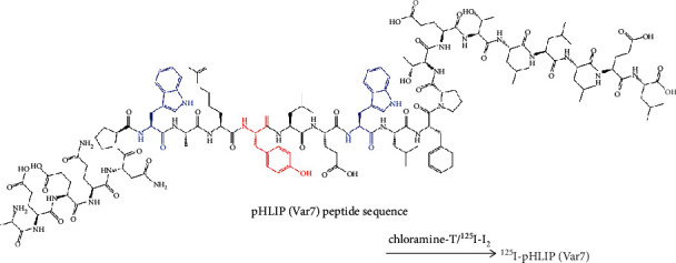
To evaluate the diagnostic efficacy of MDA-MB-231 triple-negative breast cancer with I-labeled pHLIP (Var7) by single-photon emission computed tomography/computed tomography (SPECT/CT) imaging.
The binding fraction of [I]I-pHLIP (Var7) and MDA-MB-231 cells was measured at pH 7.4 and pH 6.0, and tumor-bearing mice were subjected to small-animal SPECT/CT imaging studies.
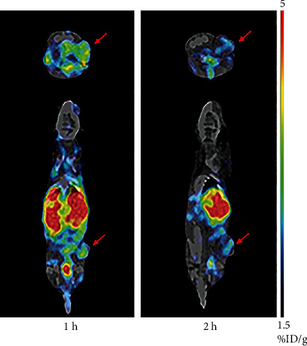
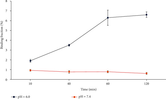
At pH = 6.0, the binding fractions of [I]I-pHLIP (Var7) and MDA-MB-231 cells at 10 min, 40 min, 1 h, and 2 h were 1.9 ± 0.1%, 3.5 ± 0.1%, 6.3 ± 0.8%, and 6.6 ± 0.3%, respectively. At pH = 7.4, there was no measured binding between [I]I-pHLIP (Var7) and MDA-MB-231 cells. Small-animal SPECT/CT imaging showed clearly visible tumors at 1 and 2 h after injection.
[I]I-pHLIP (Var7) could bind to MDA-MB-231 cells in an acidic environment, and small-animal SPECT/CT imaging showed clear tumors at 1 and 2 h after probe injection.
通过单光子发射计算机断层扫描/计算机断层扫描(SPECT/CT)成像评估 MDA-MB-231 三阴性乳腺癌的 MDA-MB-231 三阴性乳腺癌的诊断效能。
在 pH 值为 7.4 和 6.0 时测量 [I]I-pHLIP(Var7)与 MDA-MB-231 细胞的结合分数,并对荷瘤小鼠进行小动物 SPECT/CT 成像研究。
在 pH = 6.0 时,[I]I-pHLIP(Var7)与 MDA-MB-231 细胞在 10 分钟、40 分钟、1 小时和 2 小时的结合分数分别为 1.9 ± 0.1%、3.5 ± 0.1%、6.3 ± 0.8%和 6.6 ± 0.3%。在 pH = 7.4 时,[I]I-pHLIP(Var7)与 MDA-MB-231 细胞之间没有可测量的结合。小动物 SPECT/CT 成像显示在注射后 1 小时和 2 小时可以清晰地看到肿瘤。
[I]I-pHLIP(Var7)可以在酸性环境下与 MDA-MB-231 细胞结合,并且在探针注射后 1 小时和 2 小时,小动物 SPECT/CT 成像显示出清晰的肿瘤。