Wang Feng, Wei Xi-Xi, Chang Lian-Sheng, Dong Lei, Wang Yong-Ling, Li Na-Na
Henan Key Laboratory of Medical Tissue Regeneration, School of Basic Medical Sciences, Xinxiang Medical University, Xinxiang, China.
Henan Key Laboratory of Neurorestoratology (The First Affiliated Hospital of Xinxiang Medical University), Xinxiang, China.
Front Pharmacol. 2021 Mar 5;12:615104. doi: 10.3389/fphar.2021.615104. eCollection 2021.
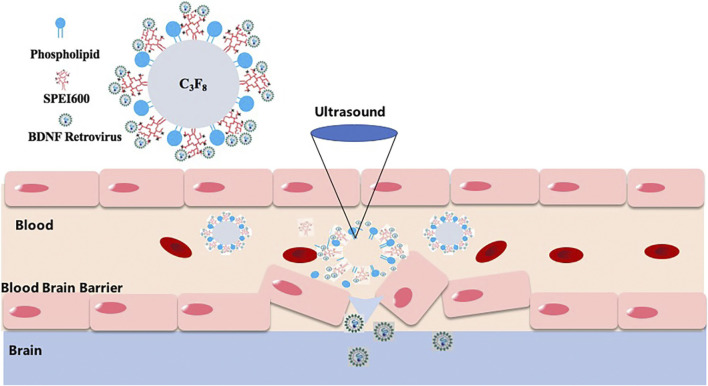
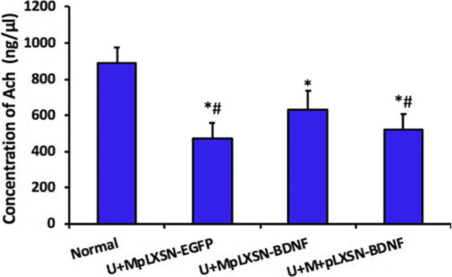
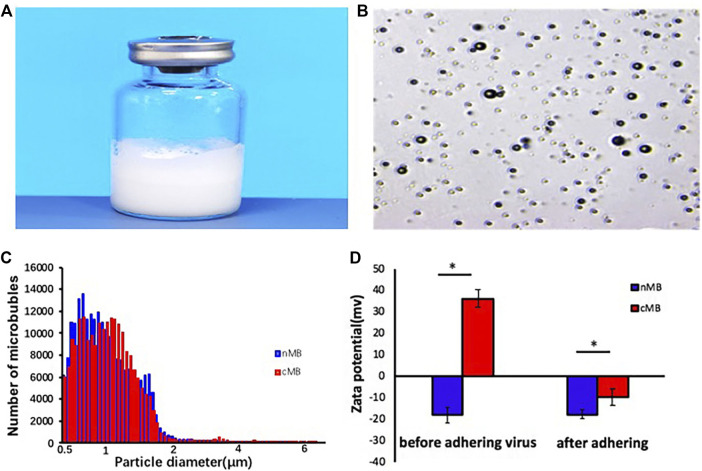
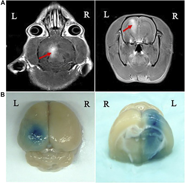
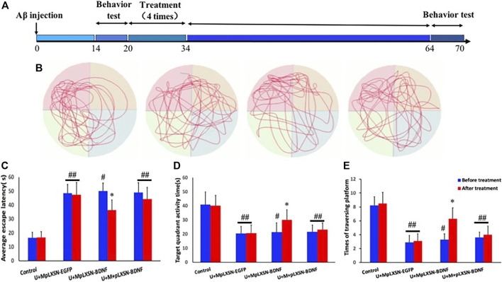
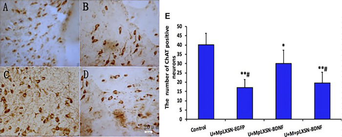
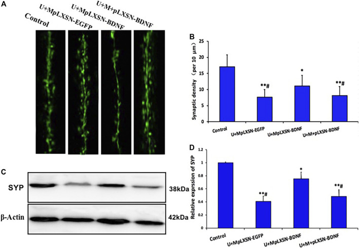
Brain-derived nerve growth factor (BDNF) is a promising effective target for the treatment of Alzheimer's disease (AD). BDNF, which has a high molecular weight, has difficulty in crossing the blood-brain barrier (BBB). The study aimed to prepare microbubbles loading brain-derived nerve growth factor (BDNF) retrovirus (MpLXSN-BDNF), to verify the characteristics of the microbubbles, and to study the therapeutic effect of the microbubbles combined with ultrasound on the opening of the blood-brain barrier in an AD rat model. 32 adult male SD rats were randomly divided into four groups: control group, ultrasound + pLXSN-EGFP microbubble group (U + MpLXSN-BDNF), ultrasound + pLXSN-BDNF microbubble group, and ultrasound + microbubble + pLXSN-BDNF virus group (U + MpLXSN-BDNF), with eight rats in each group. At the same time, the left hippocampus of rats was irradiated with low-frequency focused ultrasound guided by MRI to open the blood-brain barrier (BBB). The effects of BDNF overexpression on AD rats were evaluated behaviorally before and 1 month after the treatment. The number of acetylcholinesterase (ChAT)-positive cells and the content of acetylcholine (ACh) in brain tissues were determined by immunohistochemistry and high-performance liquid chromatography (HPLC), respectively. IF staining of synaptic spines and Western blot of synaptophysin presented herein detected synaptic density recovery. Signal intensity enhancement at the BBB disruption sites could be observed on the MR images. The behavioral evaluation showed that the times of crossing the original platform in the U + MpLXSN-BDNF group increased significantly after treatment. Immunohistochemistry and HPLC revealed that the number of ChAT-positive neurons and the contents of ACh in the brain were significantly decreased in the treated groups compared with the controls. IF staining of synaptic spines and Western blot data of synaptophysin showed that the U + MpLXSN-BDNF group can recover the synaptic loss better by BDNF supplementation than the other treatment groups. Ultrasound combined with viral microbubbles carrying BDNF can increase the transfection efficiency of brain neurons, promote the high expression of exogenous gene BDNF, and play a therapeutic role in the AD model rats.
脑源性神经营养因子(BDNF)是治疗阿尔茨海默病(AD)的一个有前景的有效靶点。BDNF分子量较大,难以穿过血脑屏障(BBB)。本研究旨在制备负载脑源性神经营养因子(BDNF)逆转录病毒的微泡(MpLXSN-BDNF),验证微泡的特性,并研究微泡联合超声对AD大鼠模型血脑屏障开放的治疗效果。32只成年雄性SD大鼠随机分为四组:对照组、超声 + pLXSN-EGFP微泡组(U + MpLXSN-BDNF)、超声 + pLXSN-BDNF微泡组和超声 + 微泡 + pLXSN-BDNF病毒组(U + MpLXSN-BDNF),每组8只。同时,在MRI引导下用低频聚焦超声照射大鼠左侧海马以开放血脑屏障(BBB)。在治疗前及治疗1个月后对AD大鼠进行行为学评估,以评价BDNF过表达对其的影响。分别通过免疫组化和高效液相色谱(HPLC)测定脑组织中乙酰胆碱酯酶(ChAT)阳性细胞数量和乙酰胆碱(ACh)含量。本文呈现的突触棘的免疫荧光染色和突触素的蛋白质印迹检测到突触密度恢复。在MR图像上可观察到血脑屏障破坏部位的信号强度增强。行为学评估显示,治疗后U + MpLXSN-BDNF组穿越原平台的次数显著增加。免疫组化和HPLC显示,与对照组相比,治疗组脑内ChAT阳性神经元数量和ACh含量显著降低。突触棘的免疫荧光染色和突触素的蛋白质印迹数据显示,U + MpLXSN-BDNF组通过补充BDNF比其他治疗组能更好地恢复突触丢失。超声联合携带BDNF的病毒微泡可提高脑神经元的转染效率,促进外源性基因BDNF的高表达,并对AD模型大鼠发挥治疗作用。