Gao Li-Juan, Dai Yuan, Li Xiao-Qiong, Meng Shi, Zhong Zhan-Qiong, Xu Shi-Jun
Institute of Meterial Medica Integration and Transformation for Brain Disorders, Chengdu University of Traditional Chinese Medicine, Chengdu, Sichuan 611137, P.R. China.
School of Pharmacy, Chengdu University of Traditional Chinese Medicine, Chengdu, Sichuan 611137, P.R. China.
Exp Ther Med. 2021 May;21(5):426. doi: 10.3892/etm.2021.9843. Epub 2021 Feb 26.
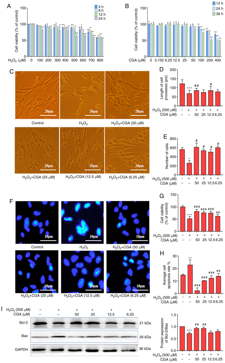
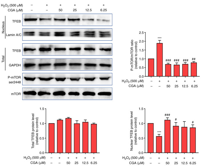
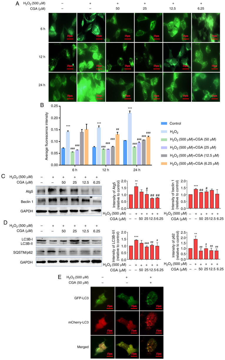
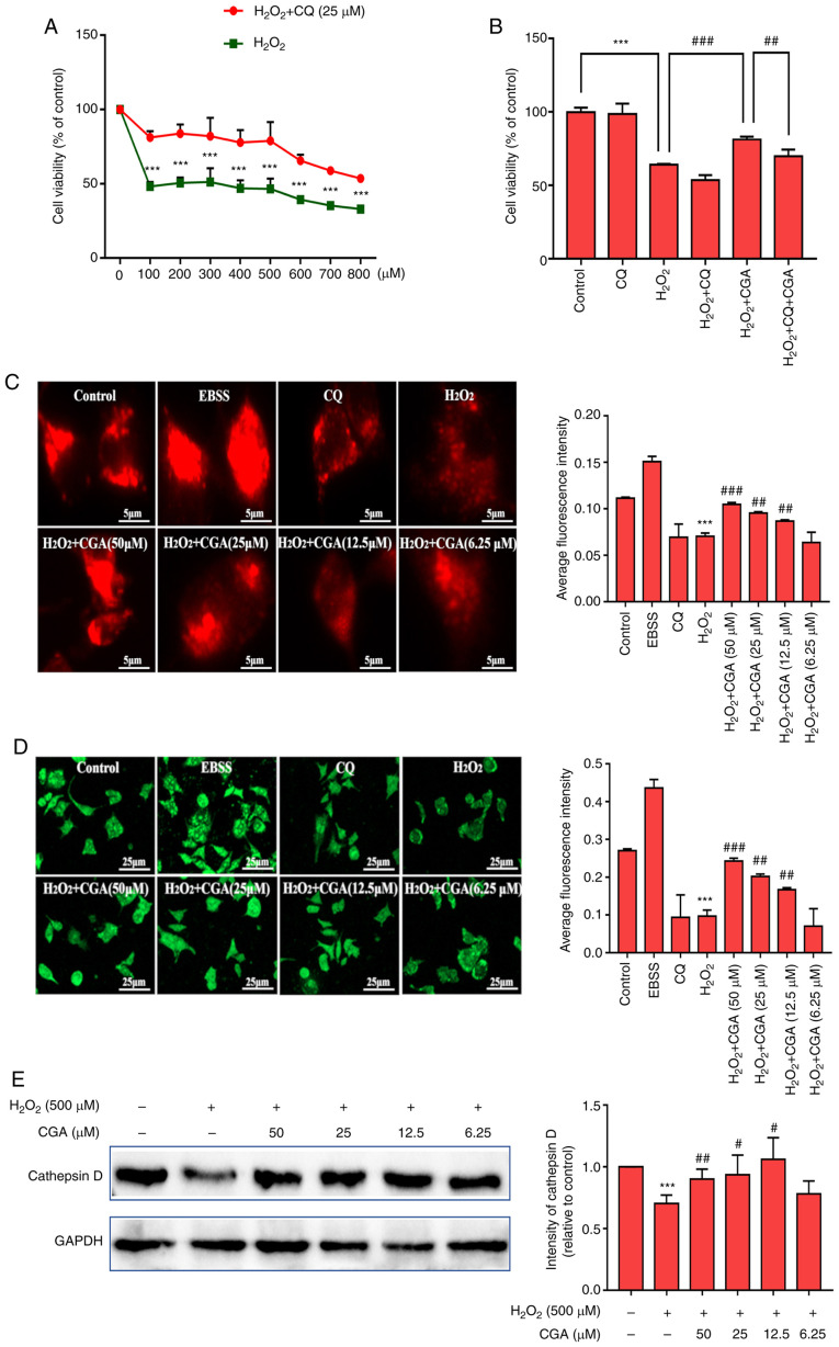
Autophagy serves an important role in amyloid-β (Aβ) metabolism and τ processing and clearance in Alzheimer's disease. The progression of Aβ plaque accumulation and hyperphosphorylation of τ proteins are enhanced by oxidative stress. A hydrogen peroxide (HO) injury cell model was established using SH-SY5Y cells. Cells were randomly divided into normal, HO and chlorogenic acid (5-caffeoylquinic acid; CGA) groups. The influence of CGA on cell viability was evaluated using a Cell Counting Kit-8 assay and cell death was assessed using Hoechst 33342 nuclear staining. Autophagy induction and fusion of autophagic vacuoles assays were performed using monodansylcadaverine staining. Additionally, SH-SY5Y cells expressing Ad-mCherry-green fluorescent protein-LC3B were established to detect autophagic flow. LysoTracker Red staining was used to evaluate lysosome function and LysoSensor™ Green staining assays were used to assess lysosomal acidification. The results demonstrated that CGA decreased the apoptosis rate, increased cell viability and improved cell morphology in HO-treated SH-SY5Y cells. Furthermore, CGA alleviated the accumulation of autophagic vacuoles, reduced the LC3BII/I ratio and decreased P62 levels, resulting in increased autophagic flux. Additionally, CGA upregulated lysosome acidity and increased the expression levels of cathepsin D. Importantly, these effects of CGA on HO-treated SH-SY5Y cells were mediated via the mTOR-transcription factor EB signaling pathway. These results indicated that CGA protected cells against HO-induced oxidative damage via the upregulation of autophagosomes, which promoted autophagocytic degradation and increased autophagic flux.
自噬在阿尔茨海默病的淀粉样β蛋白(Aβ)代谢以及τ蛋白的加工和清除过程中发挥着重要作用。氧化应激会加剧Aβ斑块的积累进程以及τ蛋白的过度磷酸化。利用SH-SY5Y细胞建立了过氧化氢(H₂O₂)损伤细胞模型。细胞被随机分为正常组、H₂O₂组和绿原酸(5-咖啡酰奎尼酸;CGA)组。使用细胞计数试剂盒-8检测法评估CGA对细胞活力的影响,并使用Hoechst 33342核染色评估细胞死亡情况。采用单丹磺酰尸胺染色进行自噬诱导和自噬泡融合检测。此外,构建了表达Ad-mCherry-绿色荧光蛋白-LC3B的SH-SY5Y细胞以检测自噬流。使用LysoTracker Red染色评估溶酶体功能,使用LysoSensor™ Green染色检测法评估溶酶体酸化。结果表明,CGA降低了H₂O₂处理的SH-SY5Y细胞的凋亡率,提高了细胞活力并改善了细胞形态。此外,CGA减轻了自噬泡的积累,降低了LC3BII/I比值并降低了P62水平,从而增加了自噬流。另外,CGA上调了溶酶体酸度并增加了组织蛋白酶D的表达水平。重要的是,CGA对H₂O₂处理的SH-SY5Y细胞的这些作用是通过mTOR-转录因子EB信号通路介导的。这些结果表明,CGA通过上调自噬体保护细胞免受H₂O₂诱导的氧化损伤,自噬体促进了自噬性降解并增加了自噬流。