Department of Histology and Embryology, School of Medicine, Yangzhou University, Yangzhou, Jiangsu 225001, P.R. China.
Mol Med Rep. 2021 May;23(5). doi: 10.3892/mmr.2021.11956. Epub 2021 Mar 24.
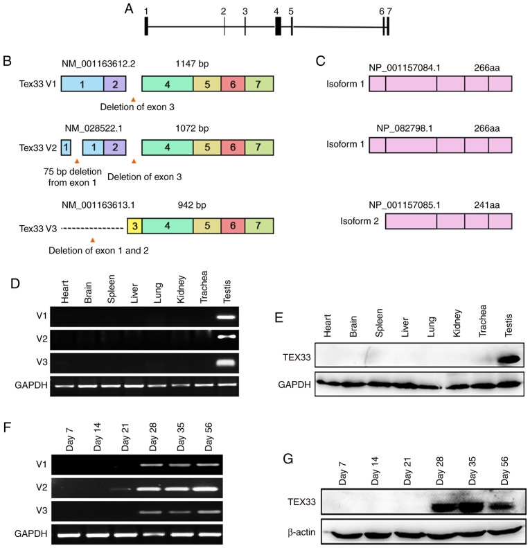
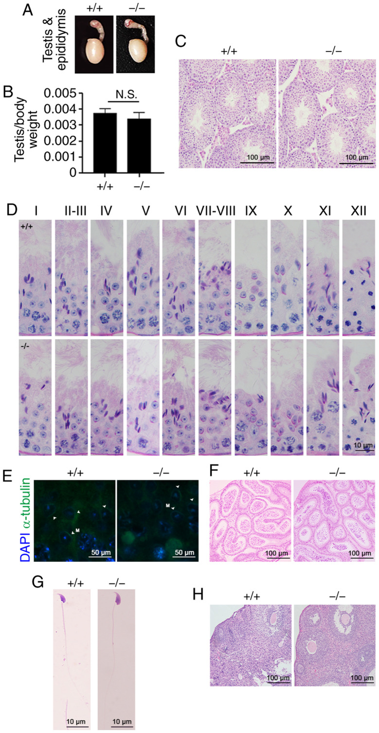
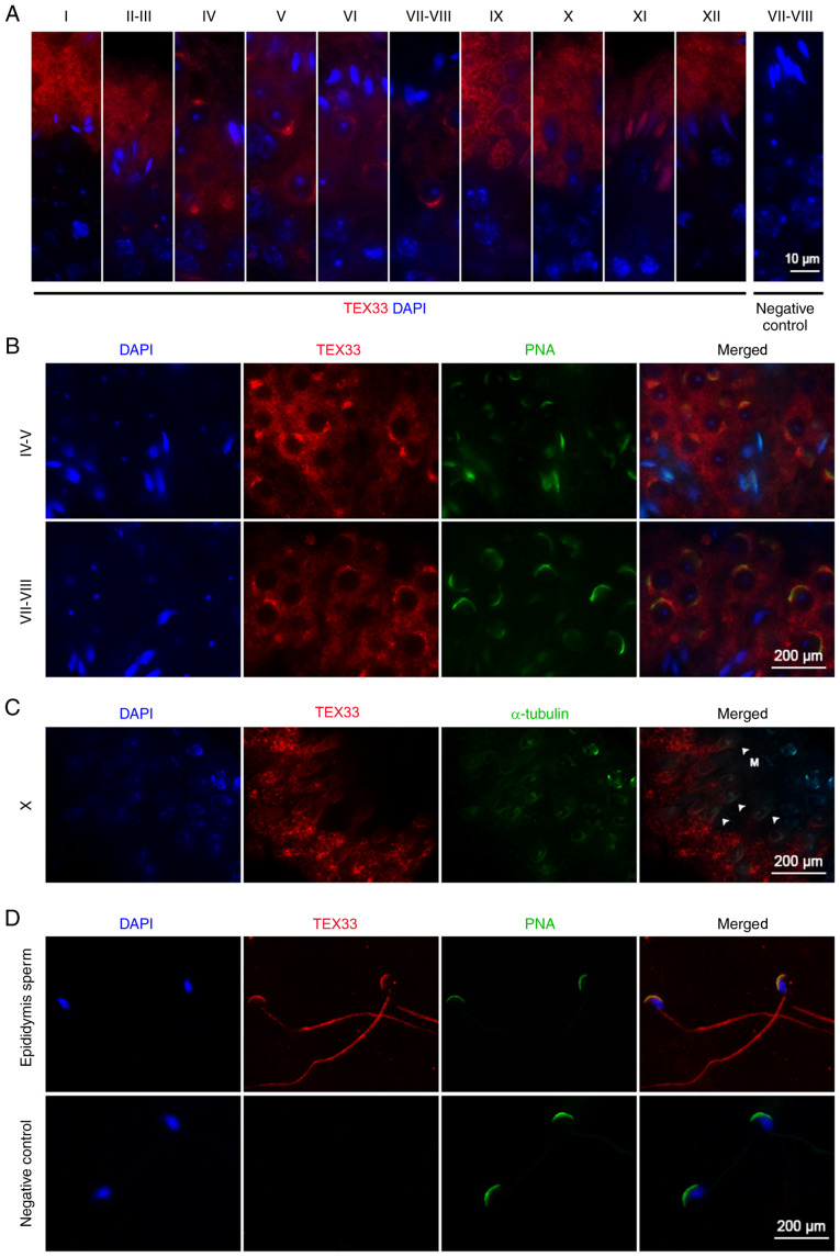
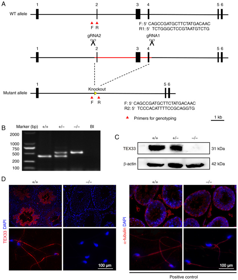
Gene expression analyses have revealed that there are >2,300 testis-enriched genes in mice, and gene knockout models have shown that a number of them are responsible for male fertility. However, the functions of numerous genes have yet to be clarified. The aim of the present study was to identify the expression pattern of testis-expressed protein 33 (TEX33) in mice and explore the role of TEX33 in male reproduction. Reverse transcription-polymerase chain reaction and western blot assays were used to investigate the mRNA and protein levels of TEX33 in mouse testes during the first wave of spermatogenesis. Immunofluorescence analysis was also performed to identify the cellular and structural localization of TEX33 protein in the testes. knockout mice were generated by CRISPR/Cas9 gene-editing. Histological analysis with hematoxylin and eosin or periodic acid-Schiff (PAS) staining, computer-assisted sperm analysis (CASA) and fertility testing, were also carried out to evaluate the effect of TEX33 on mouse spermiogenesis and male reproduction. The results showed that mRNA and protein were exclusively expressed in mouse testes and were first detected on postnatal days 21-28 (spermiogenesis phase); their expression then remained into adulthood. Immunofluorescence analysis revealed that TEX33 protein was located in the spermatids and sperm within the seminiferous tubules of the mouse testes, and exhibited specific localization to the acrosome, flagellum and manchette during spermiogenesis. These results suggested that TEX33 may play a role in mouse spermiogenesis. However, knockout mice presented no detectable difference in testis-to-body weight ratios when compared with wild-type mice. PAS staining and CASA revealed that spermatogenesis and sperm quality were normal in mice lacking . In addition, fertility testing suggested that the knockout mice had normal reproductive functions. In summary, the findings of the present study indicate that TEX33 is associated with spermiogenesis but is not essential for sperm development and male fertility.
基因表达分析表明,在小鼠中有超过 2300 个睾丸富集基因,基因敲除模型表明其中许多基因负责雄性生育能力。然而,许多基因的功能尚未阐明。本研究旨在鉴定睾丸表达蛋白 33(TEX33)在小鼠中的表达模式,并探讨 TEX33 在男性生殖中的作用。使用逆转录-聚合酶链反应和 Western blot 分析来研究第一次精子发生期间小鼠睾丸中 TEX33 的 mRNA 和蛋白水平。免疫荧光分析也用于鉴定 TEX33 蛋白在睾丸中的细胞和结构定位。通过 CRISPR/Cas9 基因编辑生成 TEX33 敲除小鼠。使用苏木精和伊红或过碘酸-希夫(PAS)染色、计算机辅助精子分析(CASA)和生育能力测试进行组织学分析,以评估 TEX33 对小鼠精子发生和雄性生殖的影响。结果表明,TEX33 mRNA 和蛋白仅在小鼠睾丸中表达,在生精期(出生后第 21-28 天)首次检测到;它们的表达随后持续到成年期。免疫荧光分析显示,TEX33 蛋白位于小鼠睾丸生精小管中的精子和精子中,在精子发生过程中特异性定位于顶体、鞭毛和生精小带。这些结果表明 TEX33 可能在小鼠精子发生中发挥作用。然而,与野生型小鼠相比,TEX33 敲除小鼠的睾丸与体重比没有明显差异。PAS 染色和 CASA 显示缺乏 的小鼠的精子发生和精子质量正常。此外,生育能力测试表明,TEX33 敲除小鼠具有正常的生殖功能。总之,本研究的结果表明 TEX33 与精子发生有关,但不是精子发育和雄性生育所必需的。