School of Biomedical Engineering and Imaging Sciences, King's College London, London, UK.
National Hospital for Neurology and Neurosurgery, London, UK.
Int J Comput Assist Radiol Surg. 2021 May;16(5):789-798. doi: 10.1007/s11548-021-02347-8. Epub 2021 Mar 24.
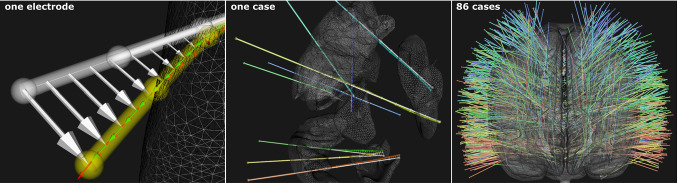
Electrode bending observed after stereotactic interventions is typically not accounted for in either computer-assisted planning algorithms, where straight trajectories are assumed, or in quality assessment, where only metrics related to entry and target points are reported. Our aim is to provide a fully automated and validated pipeline for the prediction of stereo-electroencephalography (SEEG) electrode bending. METHODS : We transform electrodes of 86 cases into a common space and compare features-based and image-based neural networks on their ability to regress local displacement ([Formula: see text]) or electrode bending ([Formula: see text]). Electrodes were stratified into six groups based on brain structures at the entry and target point. Models, both with and without Monte Carlo (MC) dropout, were trained and validated using tenfold cross-validation. RESULTS : mage-based models outperformed features-based models for all groups, and models that predicted [Formula: see text] performed better than for [Formula: see text]. Image-based model prediction with MC dropout resulted in lower mean squared error (MSE) with improvements up to 12.9% ([Formula: see text]) and 39.9% ([Formula: see text]), compared to no dropout. Using an image of brain tissue types (cortex, white and deep grey matter) resulted in similar, and sometimes better performance, compared to using a T1-weighted MRI when predicting [Formula: see text]. When inferring trajectories of image-based models (brain tissue types), 86.9% of trajectories had an MSE[Formula: see text] mm. CONCLUSION : An image-based approach regressing local displacement with an image of brain tissue types resulted in more accurate electrode bending predictions compared to other approaches, inputs, and outputs. Future work will investigate the integration of electrode bending into planning and quality assessment algorithms.
在立体定向干预后观察到的电极弯曲通常未在计算机辅助规划算法中得到考虑,因为这些算法假设了直线轨迹,或者在质量评估中,仅报告与进入点和目标点相关的指标。我们的目的是提供一种完全自动化且经过验证的流水线,用于预测立体脑电图(SEEG)电极弯曲。
我们将 86 个案例的电极转换到公共空间中,并比较基于特征和基于图像的神经网络在回归局部位移([公式:见正文])或电极弯曲([公式:见正文])方面的能力。根据进入点和目标点处的脑结构,将电极分为六组。使用十折交叉验证训练和验证具有和不具有蒙特卡罗(MC)丢弃的模型。
基于图像的模型在所有组中均优于基于特征的模型,并且预测[公式:见正文]的模型比预测[公式:见正文]的模型表现更好。与不使用 MC 丢弃相比,使用 MC 丢弃的基于图像的模型预测导致均方误差(MSE)降低,改善高达 12.9%([公式:见正文])和 39.9%([公式:见正文])。与使用 T1 加权 MRI 相比,当预测[公式:见正文]时,使用脑组织结构(皮质、白质和深部灰质)的图像会产生类似甚至更好的性能。当推断基于图像的模型(脑组织结构)的轨迹时,86.9%的轨迹的 MSE[公式:见正文]为毫米。
与其他方法、输入和输出相比,基于图像的方法通过回归脑组织结构的图像来预测局部位移,可以更准确地预测电极弯曲。未来的工作将研究将电极弯曲集成到规划和质量评估算法中的问题。