Laboratory of Chemical Pharmacology, Graduate School of Pharmaceutical Sciences, The University of Tokyo, Tokyo, Japan.
Front Immunol. 2021 Mar 8;12:617564. doi: 10.3389/fimmu.2021.617564. eCollection 2021.
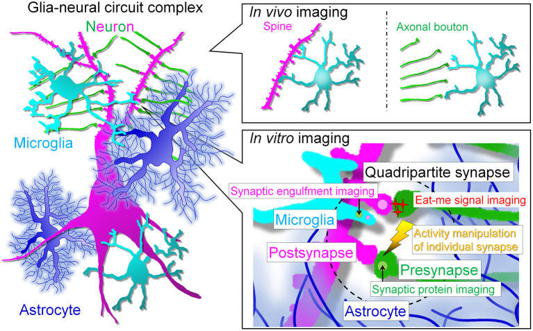
Microglia are highly dynamic in the brain in terms of their ability to migrate, proliferate, and phagocytose over the course of an individual's life. Real-time imaging is a useful tool to examine how microglial behavior is regulated and how it affects the surrounding environment. However, microglia are sensitive to environmental stimuli, so they possibly change their state during live imaging , mainly due to surgical damage, and due to various effects associated with culture conditions. Therefore, it is difficult to perform live imaging without compromising the properties of the microglia under physiological conditions. To overcome this barrier, various experimental conditions have been developed; recently, it has become possible to perform live imaging of so-called surveillant microglia , and , although there are various limitations. Now, we can choose , or live imaging systems according to the research objective. In this review, we discuss the advantages and disadvantages of each experimental system and outline the physiological significance and molecular mechanisms of microglial behavior that have been elucidated by live imaging.
小胶质细胞在其迁移、增殖和吞噬的能力方面在大脑中具有高度的动态性,贯穿个体的一生。实时成像技术是一种有用的工具,可以研究小胶质细胞的行为如何受到调节,以及它如何影响周围环境。然而,小胶质细胞对环境刺激很敏感,因此它们可能会在活体成像过程中改变状态,主要是由于手术损伤,以及与培养条件相关的各种影响。因此,在不影响小胶质细胞生理状态的情况下进行活体成像非常困难。为了克服这一障碍,已经开发了各种实验条件;最近,已经可以对所谓的“监视型”小胶质细胞进行活体成像,尽管存在各种限制。现在,我们可以根据研究目的选择活体成像系统。在这篇综述中,我们讨论了每个实验系统的优缺点,并概述了通过活体成像阐明的小胶质细胞行为的生理意义和分子机制。