F Zaky Ahmed, Froelich Michael, Meers Brad, Sturdivant Adam B, Densmore Ryan, Subramaniam Akila, Carter Tekuila, Tita Alan N, Matalon Sadis, Jilling Tamas
Anesthesiology, University of Alabama at Birmingham, Birmingham, USA.
Anesthesiology and Perioperative Medicine, University of Alabama at Birmingham School of Medicine, Birmingham, USA.
Cureus. 2021 Feb 18;13(2):e13419. doi: 10.7759/cureus.13419.
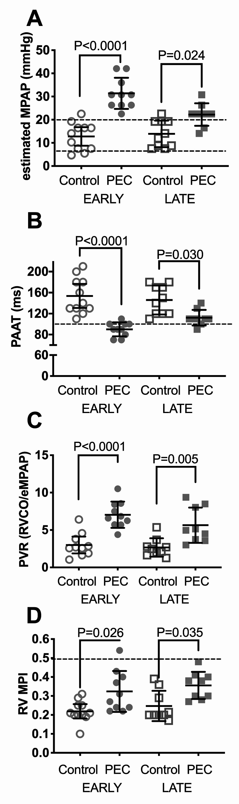
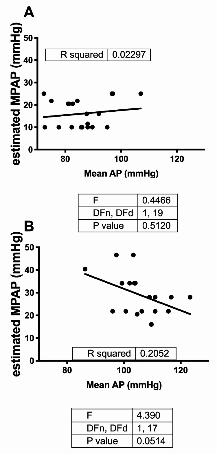
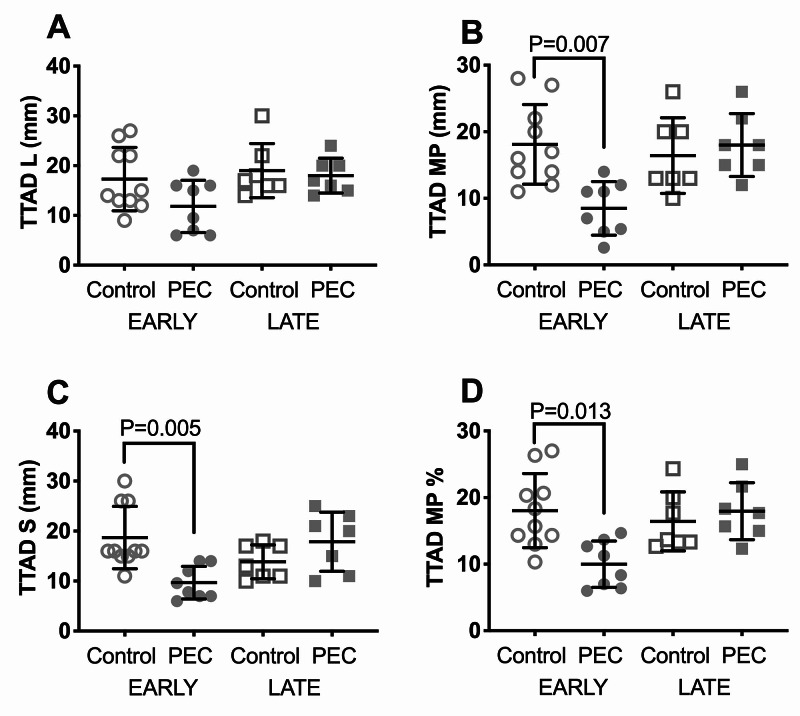
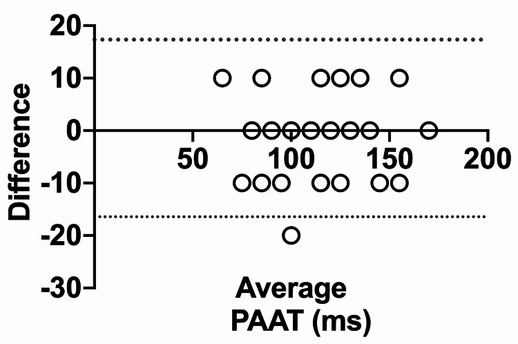
Background and objective Pre-eclampsia (PEC) is associated with the release of anti-angiogenic factors that are incriminated in raising systemic and pulmonary vascular resistance (PVR). Compared to the left heart and systemic circulation, much less attention has been paid to the right heart and pulmonary circulation in patients with PEC. We used transthoracic echocardiography (TTE) to estimate pulmonary artery (PA) pressure and right ventricular (RV) function in women with PEC. Materials and methods We conducted a case-control study at a tertiary care academic center. Ten early PEC (<34-week gestation) and nine late PEC (≥34-week gestation) patients with 11 early and 10 late gestational age-matched controls were enrolled. Two-dimensional TTE was performed on all patients. The estimated mean PA pressure (eMPAP) was calculated based on PA acceleration time (PAAT). PVR was estimated from eMPAP and RV cardiac output (RV CO). RV myocardial performance index (RV MPI), tricuspid annular plane systolic excursion (TAPSE), tissue tricuspid annular displacement (TTAD), and lateral tricuspid annular tissue peak systolic velocity (S') were measured. Results Compared to early controls, in early PEC, the eMPAP and estimated PVR (ePVR) were elevated, PAAT was reduced, RV MPI was increased, TTAD was reduced, and TAPSE and TV S' were unchanged. Compared to late controls, in late PEC, the eMPAP and ePVR were elevated, PAAT was reduced, and RV MPI was increased, while TAPSE, TTAD, and TV S' were unchanged. Conclusions In a sample of women with PEC, early PEC was found to be associated with increased eMPAP and ePVR and subclinical decrement of RV function as assessed by TTE. TTE may be a useful noninvasive screening tool for early detection of pulmonary hypertension and RV dysfunction in PEC. An adequately powered longitudinal study is needed to determine the implications of these findings on long-term outcomes.
子痫前期(PEC)与抗血管生成因子的释放有关,这些因子被认为会导致全身和肺血管阻力(PVR)升高。与左心和体循环相比,PEC患者的右心和肺循环受到的关注要少得多。我们使用经胸超声心动图(TTE)来评估PEC女性的肺动脉(PA)压力和右心室(RV)功能。
我们在一家三级医疗学术中心进行了一项病例对照研究。纳入了10例早发型PEC(孕周<34周)和9例晚发型PEC(孕周≥34周)患者,以及11例早孕期和10例晚孕期年龄匹配的对照。对所有患者进行二维TTE检查。基于肺动脉加速时间(PAAT)计算估计平均肺动脉压(eMPAP)。根据eMPAP和右心室心输出量(RV CO)估算PVR。测量右心室心肌性能指数(RV MPI)、三尖瓣环平面收缩期位移(TAPSE)、组织三尖瓣环位移(TTAD)和三尖瓣环侧壁组织峰值收缩速度(S')。
与早孕期对照组相比,早发型PEC患者的eMPAP和估计PVR(ePVR)升高,PAAT降低,RV MPI增加,TTAD降低,而TAPSE和三尖瓣S'无变化。与晚孕期对照组相比,晚发型PEC患者的eMPAP和ePVR升高,PAAT降低,RV MPI增加,而TAPSE、TTAD和三尖瓣S'无变化。
在一组PEC女性样本中,发现早发型PEC与eMPAP和ePVR升高以及通过TTE评估的右心室功能亚临床减退有关。TTE可能是早期检测PEC患者肺动脉高压和右心室功能障碍的有用无创筛查工具。需要进行一项有足够样本量的纵向研究来确定这些发现对长期结局的影响。