• 文献检索
  • 文档翻译
  • 深度研究
  • 学术资讯
  • Suppr Zotero 插件Zotero 插件
  • 邀请有礼
  • 套餐&价格
  • 历史记录
应用&插件
Suppr Zotero 插件Zotero 插件浏览器插件Mac 客户端Windows 客户端微信小程序
定价
高级版会员购买积分包购买API积分包
服务
文献检索文档翻译深度研究API 文档MCP 服务
关于我们
关于 Suppr公司介绍联系我们用户协议隐私条款
关注我们

Suppr 超能文献

核心技术专利:CN118964589B侵权必究
粤ICP备2023148730 号-1Suppr @ 2026

文献检索

告别复杂PubMed语法,用中文像聊天一样搜索,搜遍4000万医学文献。AI智能推荐,让科研检索更轻松。

立即免费搜索

文件翻译

保留排版,准确专业,支持PDF/Word/PPT等文件格式,支持 12+语言互译。

免费翻译文档

深度研究

AI帮你快速写综述,25分钟生成高质量综述,智能提取关键信息,辅助科研写作。

立即免费体验

使用基于三维超声心动图的模型将右心室划分为15个节段并分解其运动:更新后的ReVISION方法。

Partitioning the Right Ventricle Into 15 Segments and Decomposing Its Motion Using 3D Echocardiography-Based Models: The Updated ReVISION Method.

作者信息

Tokodi Márton, Staub Levente, Budai Ádám, Lakatos Bálint Károly, Csákvári Máté, Suhai Ferenc Imre, Szabó Liliána, Fábián Alexandra, Vágó Hajnalka, Tősér Zoltán, Merkely Béla, Kovács Attila

机构信息

Heart and Vascular Center, Semmelweis University, Budapest, Hungary.

Argus Cognitive, Inc., Lebanon, NH, United States.

出版信息

Front Cardiovasc Med. 2021 Mar 4;8:622118. doi: 10.3389/fcvm.2021.622118. eCollection 2021.

DOI:10.3389/fcvm.2021.622118
PMID:33763458
原文链接:https://pmc.ncbi.nlm.nih.gov/articles/PMC7982839/
Abstract

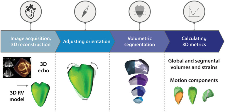
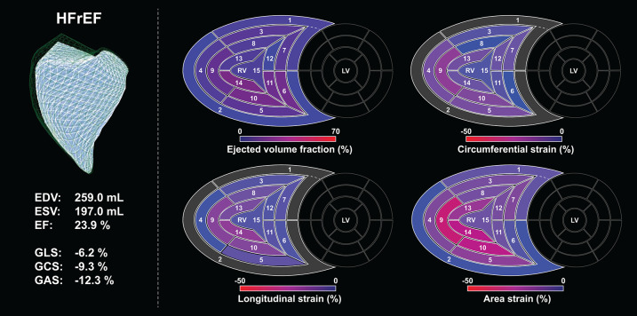
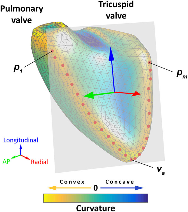
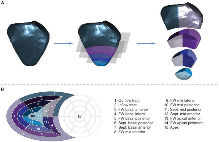
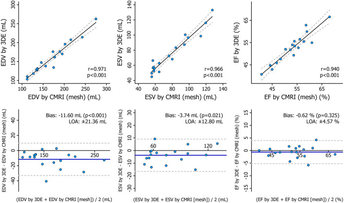
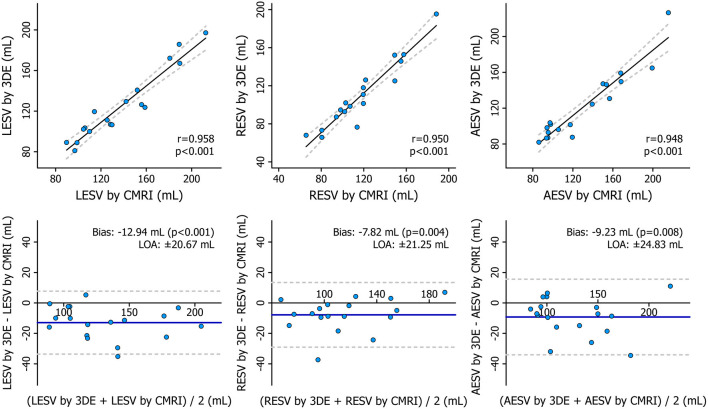
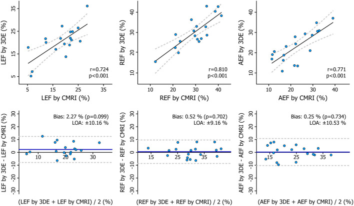
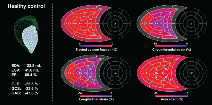
Three main mechanisms contribute to global right ventricular (RV) function: longitudinal shortening, radial displacement of the RV free wall (bellows effect), and anteroposterior shortening (as a consequence of left ventricular contraction). Since the importance of these mechanisms may vary in different cardiac conditions, a technology being able to assess their relative influence on the global RV pump function could help to clarify the pathophysiology and the mechanical adaptation of the chamber. Previously, we have introduced our 3D echocardiography (3DE)-based solution-the Right VentrIcular Separate wall motIon quantificatiON (ReVISION) method-for the quantification of the relative contribution of the three aforementioned mechanisms to global RV ejection fraction (EF). Since then, our approach has been applied in several clinical scenarios, and its strengths have been demonstrated in the in-depth characterization of RV mechanical pattern and the prognostication of patients even in the face of maintained RV EF. Recently, various new features have been implemented in our software solution to enable the convenient, standardized, and more comprehensive analysis of RV function. Accordingly, in our current technical paper, we aim to provide a detailed description of the latest version of the ReVISION method with special regards to the volumetric partitioning of the RV and the calculation of longitudinal, circumferential, and area strains using 3DE datasets. We also report the results of the comparison between 3DE- and cardiac magnetic resonance imaging-derived RV parameters, where we found a robust agreement in our advanced 3D metrics between the two modalities. In conclusion, the ReVISION method may provide novel insights into global and also segmental RV function by defining parameters that are potentially more sensitive and predictive compared to conventional echocardiographic measurements in the context of different cardiac diseases.

摘要

三种主要机制对整体右心室(RV)功能有贡献:纵向缩短、右心室游离壁的径向位移(钟摆效应)以及前后缩短(由于左心室收缩所致)。由于这些机制的重要性在不同心脏状况下可能有所不同,一种能够评估它们对整体右心室泵功能相对影响的技术有助于阐明该腔室的病理生理学和机械适应性。此前,我们已推出基于三维超声心动图(3DE)的解决方案——右心室独立壁运动量化(ReVISION)方法,用于量化上述三种机制对整体右心室射血分数(EF)的相对贡献。从那时起,我们的方法已应用于多种临床场景,并且其优势已在深入刻画右心室机械模式以及对患者进行预后评估中得到证明,即使面对右心室EF保持正常的情况也是如此。最近,我们的软件解决方案中已实现了各种新功能,以便能够对右心室功能进行便捷、标准化且更全面的分析。因此,在我们当前的技术论文中,我们旨在详细描述ReVISION方法的最新版本,特别关注右心室的容积划分以及使用3DE数据集计算纵向、圆周和面积应变。我们还报告了3DE与心脏磁共振成像得出的右心室参数之间的比较结果,我们发现这两种模式在我们先进的三维指标上具有很强的一致性。总之,ReVISION方法可能通过定义在不同心脏疾病背景下相比传统超声心动图测量可能更敏感且更具预测性的参数,为整体和节段性右心室功能提供新的见解。

https://cdn.ncbi.nlm.nih.gov/pmc/blobs/8d41/7982839/f90ffa6567b9/fcvm-08-622118-g0012.jpg
https://cdn.ncbi.nlm.nih.gov/pmc/blobs/8d41/7982839/9630b1b8cced/fcvm-08-622118-g0001.jpg
https://cdn.ncbi.nlm.nih.gov/pmc/blobs/8d41/7982839/859bdcd8e02d/fcvm-08-622118-g0002.jpg
https://cdn.ncbi.nlm.nih.gov/pmc/blobs/8d41/7982839/77ca717f8a7c/fcvm-08-622118-g0003.jpg
https://cdn.ncbi.nlm.nih.gov/pmc/blobs/8d41/7982839/d7e8b67dc614/fcvm-08-622118-g0004.jpg
https://cdn.ncbi.nlm.nih.gov/pmc/blobs/8d41/7982839/262f62296451/fcvm-08-622118-g0005.jpg
https://cdn.ncbi.nlm.nih.gov/pmc/blobs/8d41/7982839/dcb8827f9390/fcvm-08-622118-g0006.jpg
https://cdn.ncbi.nlm.nih.gov/pmc/blobs/8d41/7982839/9bc95ddd6df5/fcvm-08-622118-g0007.jpg
https://cdn.ncbi.nlm.nih.gov/pmc/blobs/8d41/7982839/8e1ca8ce8fa8/fcvm-08-622118-g0008.jpg
https://cdn.ncbi.nlm.nih.gov/pmc/blobs/8d41/7982839/0e6307ed0985/fcvm-08-622118-g0009.jpg
https://cdn.ncbi.nlm.nih.gov/pmc/blobs/8d41/7982839/81abb737a829/fcvm-08-622118-g0010.jpg
https://cdn.ncbi.nlm.nih.gov/pmc/blobs/8d41/7982839/e2e5dcca0a74/fcvm-08-622118-g0011.jpg
https://cdn.ncbi.nlm.nih.gov/pmc/blobs/8d41/7982839/f90ffa6567b9/fcvm-08-622118-g0012.jpg

相似文献

1
Partitioning the Right Ventricle Into 15 Segments and Decomposing Its Motion Using 3D Echocardiography-Based Models: The Updated ReVISION Method.使用基于三维超声心动图的模型将右心室划分为15个节段并分解其运动:更新后的ReVISION方法。
Front Cardiovasc Med. 2021 Mar 4;8:622118. doi: 10.3389/fcvm.2021.622118. eCollection 2021.
2
Quantification of the relative contribution of the different right ventricular wall motion components to right ventricular ejection fraction: the ReVISION method.不同右心室壁运动成分对右心室射血分数相对贡献的量化:ReVISION方法。
Cardiovasc Ultrasound. 2017 Mar 27;15(1):8. doi: 10.1186/s12947-017-0100-0.
3
Three-dimensional echocardiographic evaluation of longitudinal and non-longitudinal components of right ventricular contraction: results from the World Alliance of Societies of Echocardiography study.三维超声心动图评估右心室收缩的纵向和非纵向成分:来自世界超声心动图学会联盟研究的结果。
Eur Heart J Cardiovasc Imaging. 2024 Jan 29;25(2):152-160. doi: 10.1093/ehjci/jead213.
4
Importance of Nonlongitudinal Motion Components in Right Ventricular Function: Three-Dimensional Echocardiographic Study in Healthy Volunteers.右心室功能中非纵向运动分量的重要性:健康志愿者的三维超声心动图研究。
J Am Soc Echocardiogr. 2020 Aug;33(8):995-1005.e1. doi: 10.1016/j.echo.2020.04.002. Epub 2020 Jun 30.
5
Global and regional right ventricular mechanics in repaired tetralogy of Fallot with chronic severe pulmonary regurgitation: a three-dimensional echocardiography study.慢性重度肺反流的法洛四联症矫治术后右心室整体和局部心肌力学的变化:一项三维超声心动图研究。
Cardiovasc Ultrasound. 2021 Aug 6;19(1):28. doi: 10.1186/s12947-021-00260-3.
6
Global and Regional Right Ventricular Function Assessed by Novel Three-Dimensional Speckle-Tracking Echocardiography.应用新型三维斑点追踪超声心动图评估全球和区域性右心室功能。
J Am Soc Echocardiogr. 2017 Dec;30(12):1203-1213. doi: 10.1016/j.echo.2017.08.007. Epub 2017 Nov 1.
7
Accuracy and reproducibility of right ventricular quantification in patients with pressure and volume overload using single-beat three-dimensional echocardiography.使用单搏三维超声心动图对压力和容量负荷过重患者右心室定量的准确性和可重复性。
J Am Soc Echocardiogr. 2015 Mar;28(3):363-74. doi: 10.1016/j.echo.2014.10.012. Epub 2014 Dec 10.
8
A Comparison of Left- and Right-Sided Strain Software for the Assessment of Intraoperative Right Ventricular Function.左、右侧应变软件在术中右心室功能评估中的比较。
J Cardiothorac Vasc Anesth. 2019 Jun;33(6):1507-1515. doi: 10.1053/j.jvca.2018.10.038. Epub 2018 Nov 2.
9
Multi-parametric "on board" evaluation of right ventricular function using three-dimensional echocardiography: feasibility and comparison to traditional two-and three dimensional echocardiographic measurements.使用三维超声心动图对右心室功能进行多参数“术中”评估:可行性及与传统二维和三维超声心动图测量的比较
Int J Cardiovasc Imaging. 2019 Feb;35(2):275-284. doi: 10.1007/s10554-018-1496-9. Epub 2018 Nov 15.
10
Right ventricular mechanical pattern in health and disease: beyond longitudinal shortening.右心室机械模式在健康和疾病中的作用:不仅仅是纵向缩短。
Heart Fail Rev. 2019 Jul;24(4):511-520. doi: 10.1007/s10741-019-09778-1.

引用本文的文献

1
Challenges and opportunities in assessing right ventricular structure and function: a Roadmap for standardization, clinical implementation and research.评估右心室结构和功能的挑战与机遇:标准化、临床应用及研究路线图
Nat Rev Cardiol. 2025 Jun 25. doi: 10.1038/s41569-025-01180-9.
2
Impact of the right ventricular mechanical pattern assessed by three-dimensional echocardiography on adverse outcomes following cardiac surgery.三维超声心动图评估的右心室机械模式对心脏手术后不良结局的影响。
Sci Rep. 2025 Feb 15;15(1):5623. doi: 10.1038/s41598-025-89122-w.
3
Three-Dimensional Echocardiographic Assessment of Right Ventricular Global Myocardial Work and Ventricular-Pulmonary Coupling in ATTR Cardiac Amyloidosis.

本文引用的文献

1
Volumetric parcellation of the cardiac right ventricle for regional geometric and functional assessment.心脏右心室容积分割用于区域性几何和功能评估。
Med Image Anal. 2021 Jul;71:102044. doi: 10.1016/j.media.2021.102044. Epub 2021 Apr 6.
2
Importance of Nonlongitudinal Motion Components in Right Ventricular Function: Three-Dimensional Echocardiographic Study in Healthy Volunteers.右心室功能中非纵向运动分量的重要性:健康志愿者的三维超声心动图研究。
J Am Soc Echocardiogr. 2020 Aug;33(8):995-1005.e1. doi: 10.1016/j.echo.2020.04.002. Epub 2020 Jun 30.
3
Right ventricular three-dimensional echocardiography: the current status and future perspectives.
转甲状腺素蛋白型心脏淀粉样变中右心室整体心肌做功及心室-肺耦联的三维超声心动图评估
J Clin Med. 2025 Jan 21;14(3):668. doi: 10.3390/jcm14030668.
4
Pathophysiology of the right ventricle and its pulmonary vascular interaction.右心室的病理生理学及其与肺血管的相互作用。
Eur Respir J. 2024 Oct 31;64(4). doi: 10.1183/13993003.01321-2024. Print 2024 Oct.
5
The effects of mitral stenosis on right ventricular mechanics assessed by three-dimensional echocardiography.三维超声心动图评估二尖瓣狭窄对右心室力学的影响。
Sci Rep. 2024 Jul 24;14(1):17112. doi: 10.1038/s41598-024-68126-y.
6
Right Ventricular Structure and Function in Adolescent Athletes: A 3D Echocardiographic Study.青少年运动员的右心室结构和功能:一项 3D 超声心动图研究。
Int J Sports Med. 2024 Jun;45(6):473-480. doi: 10.1055/a-2259-2203. Epub 2024 Feb 1.
7
Editorial: Added value of 3D imaging in the diagnosis and prognostication of patients with right ventricular dysfunction.社论:三维成像在右心室功能障碍患者诊断和预后评估中的附加价值
Front Cardiovasc Med. 2024 Jan 9;10:1356294. doi: 10.3389/fcvm.2023.1356294. eCollection 2023.
8
Added value of 3D echocardiography in the diagnosis and prognostication of patients with right ventricular dysfunction.三维超声心动图在右心室功能障碍患者诊断及预后评估中的附加价值
Front Cardiovasc Med. 2023 Dec 21;10:1263864. doi: 10.3389/fcvm.2023.1263864. eCollection 2023.
9
Perioperative Quality Initiative (POQI) consensus statement on perioperative assessment of right ventricular function.围手术期质量改进计划(POQI)关于右心室功能围手术期评估的共识声明。
Perioper Med (Lond). 2023 Dec 8;12(1):66. doi: 10.1186/s13741-023-00351-x.
10
Right ventricular contraction patterns in healthy children using three-dimensional echocardiography.使用三维超声心动图观察健康儿童的右心室收缩模式。
Front Cardiovasc Med. 2023 Aug 3;10:1141027. doi: 10.3389/fcvm.2023.1141027. eCollection 2023.
右心室三维超声心动图:现状与未来展望。
J Echocardiogr. 2020 Sep;18(3):149-159. doi: 10.1007/s12574-020-00468-8. Epub 2020 Apr 2.
4
Right ventricular mechanical pattern in patients undergoing mitral valve surgery: a predictor of post-operative dysfunction?二尖瓣手术患者的右心室机械模式:术后功能障碍的预测因子?
ESC Heart Fail. 2020 Jun;7(3):1246-1256. doi: 10.1002/ehf2.12682. Epub 2020 Mar 26.
5
Arrhythmogenic right ventricular cardiomyopathy: evaluation of the current diagnostic criteria and differential diagnosis.致心律失常性右室心肌病:当前诊断标准及鉴别诊断的评估
Eur Heart J. 2020 Apr 7;41(14):1414-1429. doi: 10.1093/eurheartj/ehz669.
6
Relative Prognostic Importance of Left and Right Ventricular Ejection Fraction in Patients With Cardiac Diseases.左、右心室射血分数对心脏病患者预后的相对重要性。
J Am Soc Echocardiogr. 2019 Nov;32(11):1407-1415.e3. doi: 10.1016/j.echo.2019.06.009. Epub 2019 Aug 7.
7
Right ventricular mechanical pattern in health and disease: beyond longitudinal shortening.右心室机械模式在健康和疾病中的作用:不仅仅是纵向缩短。
Heart Fail Rev. 2019 Jul;24(4):511-520. doi: 10.1007/s10741-019-09778-1.
8
Characterization of Right Ventricular Deformation in Pulmonary Arterial Hypertension Using Three-Dimensional Principal Strain Analysis.使用三维主应变分析技术对肺动脉高压患者右心室变形进行特征描述。
J Am Soc Echocardiogr. 2019 Mar;32(3):385-393. doi: 10.1016/j.echo.2018.10.001. Epub 2018 Dec 11.
9
Characterization of the dynamic changes in left ventricular morphology and function induced by exercise training and detraining.运动训练和停训诱导左心室形态和功能动态变化的特征。
Int J Cardiol. 2019 Feb 15;277:178-185. doi: 10.1016/j.ijcard.2018.10.092. Epub 2018 Nov 1.
10
Cardiac magnetic resonance based deformation imaging: role of feature tracking in athletes with suspected arrhythmogenic right ventricular cardiomyopathy.基于心脏磁共振的形变成像:特征追踪在疑似致心律失常性右室心肌病运动员中的作用
Int J Cardiovasc Imaging. 2019 Mar;35(3):529-538. doi: 10.1007/s10554-018-1478-y. Epub 2018 Oct 31.