Post-Graduation Program of Dentistry, Department of Orthodontics, Federal University of Pará, Belém, Pará, Brazil.
Dental School, Federal University of Pará, Belém, Pará, Brazil.
PLoS One. 2021 Mar 25;16(3):e0249119. doi: 10.1371/journal.pone.0249119. eCollection 2021.
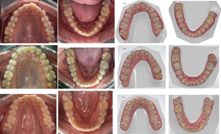
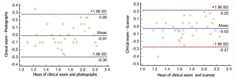
Dental wear analysis through the use of an intraoral scanner is a reality of modern dentistry. This study aimed to investigate the reliability of qualitative tooth wear evaluation through three-dimensional images captured with an intraoral scanner and compared to clinical and photographic examinations. Eighteen adult volunteers of both genders (18 to 55 years old) were submitted to clinical exams, intraoral photographs and intraoral scanning protocol using an optical scanner (TRIOS® Pod, 3Shape, Copenhagen, Denmark). Occlusal tooth wear, from second to second premolars, was measured by two evaluators and reevaluated after 30 days, according to a slight modification of the method described by Mockers et al. Weighted Kappa was used to measure intra and inter-examiner agreement. The Friedman test was used to verify the differences among methods. Random and systematic errors were assessed using Bland-Altman plots. All statistical analysis was performed with p<0.05. There was a substantive agreement for clinical (K = 0.75) and photographic exams (K = 0.79) and a moderate agreement for intraoral scanner analysis (K = 0.60) for inter-examiner evaluation. A substantial intra-examiner agreement was obtained for both evaluators. No significant difference between the methods was observed (p = 0.7343 for examiner 1 and 0.8007 for examiner 2). The Bland-Altman plot confirmed no systematic errors between the methods and a random error of 0.25 with the scanner method when compared to clinical assessment. All three methods showed reliability in qualitative occlusal tooth wear evaluation. Intraoral scanning seems to be a sound and reliable tool to evaluate tooth wear when compared to traditional methods, considering the lower inter-examiner agreement and the inherent limitations of this pilot study. Further research will be necessary in order to achieve more robust evidence.
通过使用口腔内扫描仪进行牙齿磨损分析是现代牙科的现实。本研究旨在通过三维图像评估定性牙齿磨损的可靠性,这些图像是使用口腔内扫描仪拍摄的,并与临床和摄影检查进行比较。18 名成年志愿者(18 至 55 岁)接受了临床检查、口腔内照片和光学扫描仪(TRIOS® Pod,3Shape,哥本哈根,丹麦)的口腔内扫描方案。根据 Mockers 等人描述的方法进行了轻微修改,由两位评估者评估从第二前磨牙到第二前磨牙的咬合面牙齿磨损,并在 30 天后重新评估。使用加权 Kappa 评估评估者内和评估者间的一致性。使用 Friedman 检验验证方法之间的差异。使用 Bland-Altman 图评估随机和系统误差。所有统计分析均采用 p<0.05。临床检查(K = 0.75)和摄影检查(K = 0.79)的评估者间有实质性一致性,口腔内扫描仪分析(K = 0.60)的评估者间有中度一致性。两位评估者都获得了实质性的评估者内一致性。方法之间没有观察到显著差异(评估者 1 为 p = 0.7343,评估者 2 为 0.8007)。Bland-Altman 图证实方法之间没有系统误差,与临床评估相比,扫描仪方法的随机误差为 0.25。所有三种方法在定性咬合面牙齿磨损评估中均具有可靠性。与传统方法相比,口腔内扫描似乎是一种可靠的工具,考虑到评估者间的一致性较低,以及本研究的固有局限性,还需要进一步的研究来获得更可靠的证据。