Department of Immunology and Pathology, Central Clinical School, Monash University, Melbourne, VIC, Australia.
Department of Allergy, Immunology and Respiratory Medicine, Central Clinical School, Monash University, Melbourne, VIC, Australia.
Allergy. 2021 Oct;76(10):3028-3040. doi: 10.1111/all.14832. Epub 2021 May 5.
Diagnostic tests for allergy rely on detecting allergen-specific IgE. Component-resolved diagnostics incorporate multiple defined allergen components to improve the quality of diagnosis and patient care.
To develop a new approach for determining sensitization to specific allergen components that utilizes fluorescent protein tetramers for direct staining of IgE on blood basophils by flow cytometry.
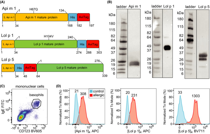
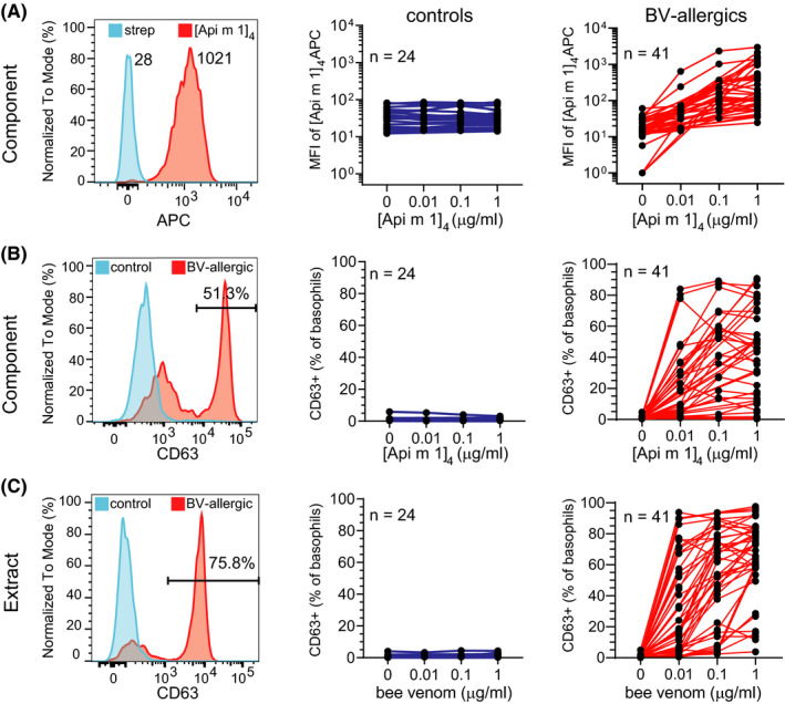
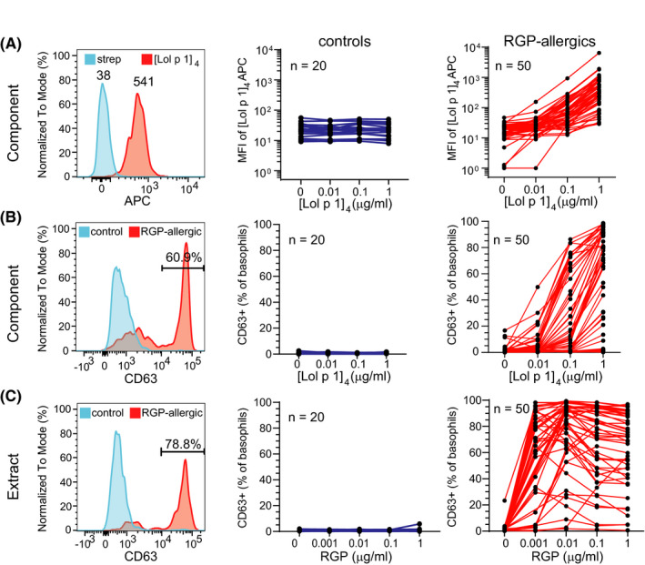
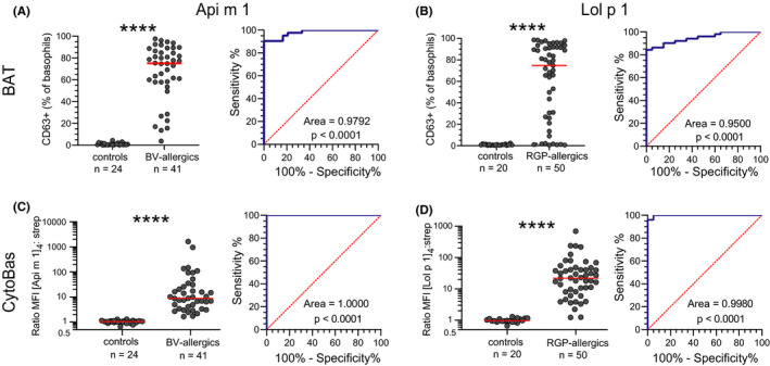
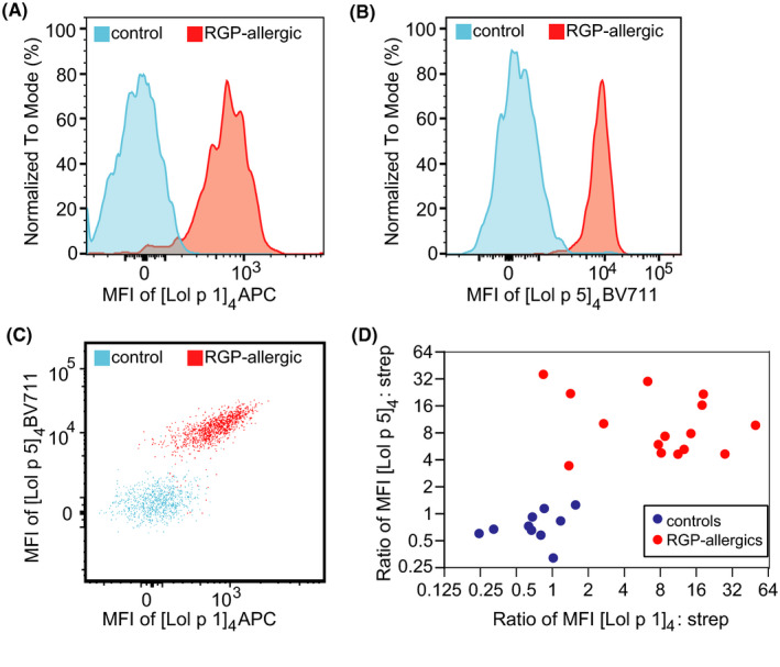
Recombinant forms of Lol p 1 and Lol p 5 proteins from ryegrass pollen (RGP) and Api m 1 from honeybee venom (BV) were produced, biotinylated, and tetramerized with streptavidin-fluorochrome conjugates. Blood samples from 50 RGP-allergic, 41 BV-allergic, and 26 controls were incubated with fluorescent protein tetramers for flow cytometric evaluation of basophil allergen binding and activation.
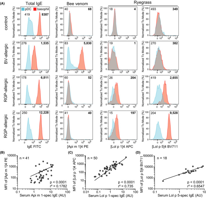
Allergen tetramers bound to and activated basophils from relevant allergic patients but not controls. Direct fluorescence staining of Api m 1 and Lol p 1 tetramers had greater positive predictive values than basophil activation for BV and RGP allergy, respectively, as defined with receiver operator characteristics (ROC) curves. Staining intensities of allergen tetramers correlated with allergen-specific IgE levels in serum. Inclusion of multiple allergens coupled with distinct fluorochromes in a single-tube assay enabled rapid detection of sensitization to both Lol p 1 and Lol p 5 in RGP-allergic patients and discriminated between controls, BV-allergic, and RGP-allergic patients.
Our novel flow cytometric assay, termed CytoBas, enables rapid and reliable detection of clinically relevant allergic sensitization. The intensity of fluorescent allergen tetramer staining of basophils has a high positive predictive value for disease, and the assay can be multiplexed for a component-resolved and differential diagnostic test for allergy.
过敏的诊断测试依赖于检测过敏原特异性 IgE。成分解析诊断结合了多个定义明确的过敏原成分,以提高诊断和患者护理的质量。
开发一种新方法,用于确定对特定过敏原成分的致敏性,该方法利用荧光蛋白四聚体通过流式细胞术直接染色血液嗜碱性粒细胞上的 IgE。
从黑麦草花粉(RGP)中产生重组 Lol p 1 和 Lol p 5 蛋白形式,从蜂毒液(BV)中产生 Api m 1 蛋白形式,然后用生物素化和链霉亲和素-荧光染料缀合物四聚化。将 50 名 RGP 过敏、41 名 BV 过敏和 26 名对照的血液样本与荧光蛋白四聚体孵育,通过流式细胞术评估嗜碱性粒细胞过敏原结合和激活。
过敏原四聚体与相关过敏患者的嗜碱性粒细胞结合并激活,但与对照者不结合。使用接收器操作特征(ROC)曲线定义,直接荧光染色 Api m 1 和 Lol p 1 四聚体对 BV 和 RGP 过敏的阳性预测值分别大于嗜碱性粒细胞激活。过敏原四聚体的染色强度与血清中过敏原特异性 IgE 水平相关。在单个试管测定中包含多种过敏原并结合不同的荧光染料,可快速检测 RGP 过敏患者对 Lol p 1 和 Lol p 5 的致敏,并区分对照者、BV 过敏者和 RGP 过敏者。
我们的新型流式细胞术测定法,称为 CytoBas,可快速可靠地检测临床相关的过敏致敏。嗜碱性粒细胞中荧光过敏原四聚体染色的强度对疾病具有高阳性预测值,并且该测定法可进行多重分析,用于过敏的成分解析和鉴别诊断测试。