• 文献检索
  • 文档翻译
  • 深度研究
  • 学术资讯
  • Suppr Zotero 插件Zotero 插件
  • 邀请有礼
  • 套餐&价格
  • 历史记录
应用&插件
Suppr Zotero 插件Zotero 插件浏览器插件Mac 客户端Windows 客户端微信小程序
定价
高级版会员购买积分包购买API积分包
服务
文献检索文档翻译深度研究API 文档MCP 服务
关于我们
关于 Suppr公司介绍联系我们用户协议隐私条款
关注我们

Suppr 超能文献

核心技术专利:CN118964589B侵权必究
粤ICP备2023148730 号-1Suppr @ 2026

文献检索

告别复杂PubMed语法,用中文像聊天一样搜索,搜遍4000万医学文献。AI智能推荐,让科研检索更轻松。

立即免费搜索

文件翻译

保留排版,准确专业,支持PDF/Word/PPT等文件格式,支持 12+语言互译。

免费翻译文档

深度研究

AI帮你快速写综述,25分钟生成高质量综述,智能提取关键信息,辅助科研写作。

立即免费体验

单细胞肝脏 T 细胞图谱揭示原发性硬化性胆管炎中肝固有初始样 CD4 T 细胞的扩增。

Single-cell atlas of hepatic T cells reveals expansion of liver-resident naive-like CD4 T cells in primary sclerosing cholangitis.

机构信息

I. Department of Medicine, University Medical Center Hamburg-Eppendorf, Hamburg 20246 Germany.

I. Department of Medicine, University Medical Center Hamburg-Eppendorf, Hamburg 20246 Germany; Bioinformatics Core, University Medical Center Hamburg-Eppendorf, Hamburg 20246 Germany.

出版信息

J Hepatol. 2021 Aug;75(2):414-423. doi: 10.1016/j.jhep.2021.03.016. Epub 2021 Mar 24.

DOI:10.1016/j.jhep.2021.03.016
PMID:33774059
原文链接:https://pmc.ncbi.nlm.nih.gov/articles/PMC8310924/
Abstract

BACKGROUND & AIMS: Little is known about the composition of intrahepatic immune cells and their contribution to the pathogenesis of primary sclerosing cholangitis (PSC). Herein, we aimed to create an atlas of intrahepatic T cells and thereby perform an in-depth characterization of T cells in inflamed human liver.

METHODS

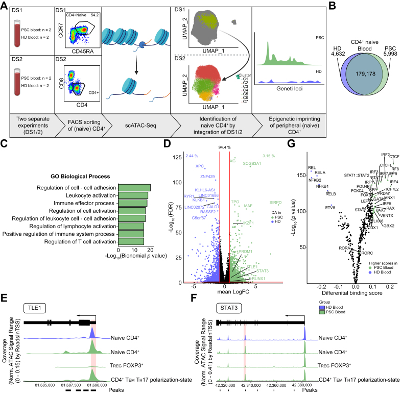
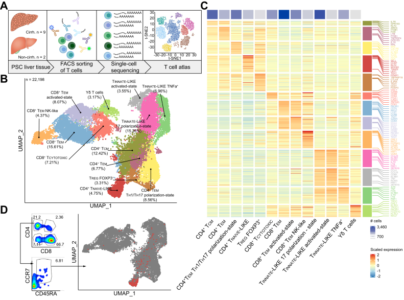
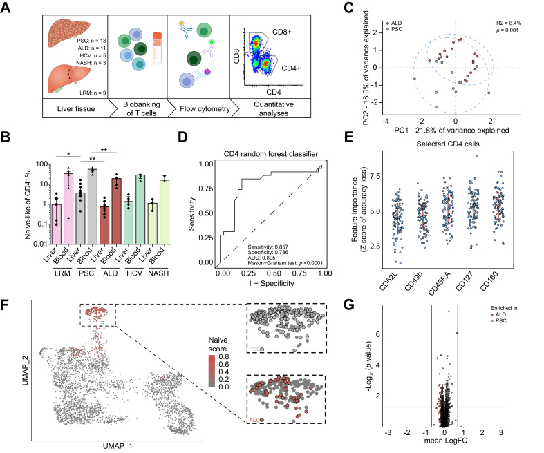
Different single-cell RNA sequencing methods were combined with in silico analyses on intrahepatic and peripheral T cells from patients with PSC (n = 11) and healthy donors (HDs, n = 4). Multi-parameter flow cytometry and functional in vitro experiments were conducted on samples from patients with PSC (n = 24), controls with other liver diseases and HDs.

RESULTS

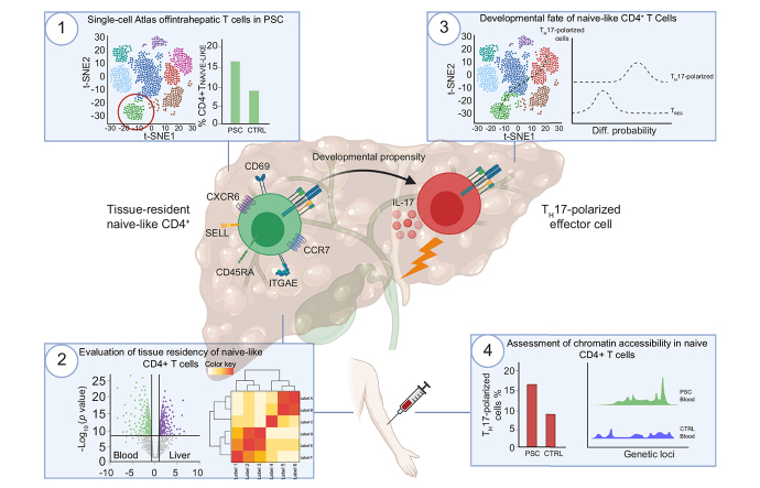
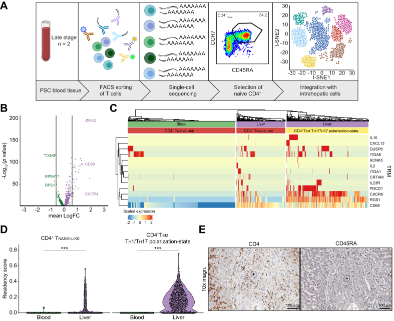
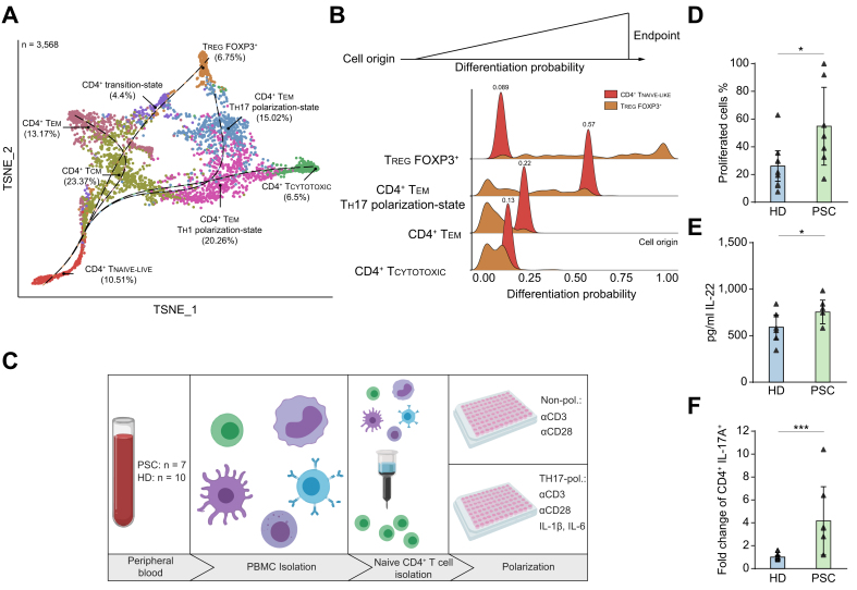
We identified a population of intrahepatic naive-like CD4 T cells, which was present in all liver diseases tested, but particularly expanded in PSC. This population had a transcriptome and T cell receptor repertoire similar to circulating naive T cells but expressed a set of genes associated with tissue residency. Their periductal location supported the concept of tissue-resident naive-like T cells in livers of patients with PSC. Trajectory inference suggested that these cells had the developmental propensity to acquire a T helper 17 (T17) polarization state. Functional and chromatin accessibility experiments revealed that circulating naive T cells in patients with PSC were predisposed to polarize towards T17 cells.

CONCLUSION

We report the first atlas of intrahepatic T cells in PSC, which led to the identification of a previously unrecognized population of tissue-resident naive-like T cells in the inflamed human liver and to the finding that naive CD4 T cells in PSC harbour the propensity to develop into T17 cells.

LAY SUMMARY

The composition of intrahepatic immune cells in primary sclerosing cholangitis (PSC) and their contribution to disease pathogenesis is widely unknown. We analysed intrahepatic T cells and identified a previously uncharacterized population of liver-resident CD4 T cells which are expanded in the livers of patients with PSC compared to healthy liver tissue and other liver diseases. These cells are likely to contribute to the pathogenesis of PSC and could be targeted in novel therapeutic approaches.

摘要

背景与目的

对于肝内免疫细胞的组成及其在原发性硬化性胆管炎(PSC)发病机制中的作用知之甚少。在此,我们旨在创建肝内 T 细胞图谱,从而深入分析炎症性人类肝脏中的 T 细胞。

方法

我们结合了不同的单细胞 RNA 测序方法,并对来自 PSC 患者(n=11)和健康供体(HD,n=4)的肝内和外周 T 细胞进行了计算机分析。我们对来自 PSC 患者(n=24)、其他肝病患者和 HD 的样本进行了多参数流式细胞术和功能体外实验。

结果

我们鉴定了一群肝内初始样 CD4 T 细胞,该细胞存在于所有测试的肝脏疾病中,但在 PSC 中特别扩增。该细胞群具有与循环初始 T 细胞相似的转录组和 T 细胞受体库,但表达了一组与组织驻留相关的基因。它们位于胆管周围的位置支持 PSC 患者肝脏中存在组织驻留初始样 T 细胞的概念。轨迹推断表明,这些细胞具有获得 T 辅助 17(T17)极化状态的发育倾向。功能和染色质可及性实验表明,PSC 患者的循环初始 T 细胞倾向于向 T17 细胞极化。

结论

我们报告了 PSC 中肝内 T 细胞的首个图谱,该图谱导致鉴定出了在炎症性人类肝脏中以前未被识别的组织驻留初始样 T 细胞群,并发现 PSC 中的初始 CD4 T 细胞具有发展为 T17 细胞的倾向。

概要

原发性硬化性胆管炎(PSC)肝内免疫细胞的组成及其对疾病发病机制的贡献尚不清楚。我们分析了肝内 T 细胞,鉴定了一群以前未被描述的肝驻留 CD4 T 细胞,与健康肝组织和其他肝病相比,这些细胞在 PSC 患者的肝脏中扩增。这些细胞可能有助于 PSC 的发病机制,并且可以作为新型治疗方法的靶点。

https://cdn.ncbi.nlm.nih.gov/pmc/blobs/99cf/8310924/670e34d6d6ee/gr5.jpg
https://cdn.ncbi.nlm.nih.gov/pmc/blobs/99cf/8310924/3121433e4f08/ga1.jpg
https://cdn.ncbi.nlm.nih.gov/pmc/blobs/99cf/8310924/ff33a0e17f16/gr1.jpg
https://cdn.ncbi.nlm.nih.gov/pmc/blobs/99cf/8310924/28716399c593/gr2.jpg
https://cdn.ncbi.nlm.nih.gov/pmc/blobs/99cf/8310924/fd4f00cacfc3/gr3.jpg
https://cdn.ncbi.nlm.nih.gov/pmc/blobs/99cf/8310924/db4b265cc616/gr4.jpg
https://cdn.ncbi.nlm.nih.gov/pmc/blobs/99cf/8310924/670e34d6d6ee/gr5.jpg

相似文献

1
Single-cell atlas of hepatic T cells reveals expansion of liver-resident naive-like CD4 T cells in primary sclerosing cholangitis.单细胞肝脏 T 细胞图谱揭示原发性硬化性胆管炎中肝固有初始样 CD4 T 细胞的扩增。
J Hepatol. 2021 Aug;75(2):414-423. doi: 10.1016/j.jhep.2021.03.016. Epub 2021 Mar 24.
2
Single-cell, single-nucleus, and spatial transcriptomics characterization of the immunological landscape in the healthy and PSC human liver.单细胞、单细胞核和空间转录组学分析健康和 PSC 人肝中的免疫景观。
J Hepatol. 2024 May;80(5):730-743. doi: 10.1016/j.jhep.2023.12.023. Epub 2024 Jan 8.
3
Interferon-γ-dependent immune responses contribute to the pathogenesis of sclerosing cholangitis in mice.干扰素-γ 依赖性免疫应答有助于小鼠硬化性胆管炎的发病机制。
J Hepatol. 2019 Oct;71(4):773-782. doi: 10.1016/j.jhep.2019.05.023. Epub 2019 Jun 5.
4
Gut and liver T-cells of common clonal origin in primary sclerosing cholangitis-inflammatory bowel disease.原发性硬化性胆管炎-炎症性肠病中共同克隆起源的肠道和肝脏 T 细胞。
J Hepatol. 2017 Jan;66(1):116-122. doi: 10.1016/j.jhep.2016.09.002. Epub 2016 Sep 17.
5
Downregulation of TGR5 (GPBAR1) in biliary epithelial cells contributes to the pathogenesis of sclerosing cholangitis.TGR5(GPBAR1)在胆管上皮细胞中的下调导致硬化性胆管炎的发病机制。
J Hepatol. 2021 Sep;75(3):634-646. doi: 10.1016/j.jhep.2021.03.029. Epub 2021 Apr 17.
6
Inflammatory type 2 conventional dendritic cells contribute to murine and human cholangitis.炎症型 2 型常规树突状细胞有助于引发鼠类和人类胆管炎。
J Hepatol. 2022 Dec;77(6):1532-1544. doi: 10.1016/j.jhep.2022.06.025. Epub 2022 Jul 5.
7
Macrophages contribute to the pathogenesis of sclerosing cholangitis in mice.巨噬细胞促进了小鼠硬化性胆管炎的发病机制。
J Hepatol. 2018 Sep;69(3):676-686. doi: 10.1016/j.jhep.2018.05.018. Epub 2018 May 24.
8
Intergenic risk variant rs56258221 skews the fate of naive CD4 T cells via miR4464-BACH2 interplay in primary sclerosing cholangitis.基因间风险变异rs56258221通过原发性硬化性胆管炎中miR4464与BACH2的相互作用影响初始CD4 T细胞的命运。
Cell Rep Med. 2024 Jul 16;5(7):101620. doi: 10.1016/j.xcrm.2024.101620. Epub 2024 Jun 19.
9
Dysfunction of hepatic regulatory T cells in experimental sclerosing cholangitis is related to IL-12 signaling.实验性硬化性胆管炎中肝调节性 T 细胞功能障碍与 IL-12 信号有关。
J Hepatol. 2017 Apr;66(4):798-805. doi: 10.1016/j.jhep.2016.12.001. Epub 2016 Dec 10.
10
Itaconate inhibits CD103 + T RM cells and alleviates hepatobiliary injury in mouse models of primary sclerosing cholangitis.衣康酸酯抑制原发性硬化性胆管炎小鼠模型中的CD103 + 组织驻留记忆T细胞并减轻肝胆损伤。
Hepatology. 2024 Jan 1;79(1):25-38. doi: 10.1097/HEP.0000000000000549. Epub 2023 Jul 26.

引用本文的文献

1
Macrophages drive a fibrogenic gene program of periductal fibroblasts in pediatric primary sclerosing cholangitis.巨噬细胞驱动小儿原发性硬化性胆管炎中导管周围成纤维细胞的促纤维化基因程序。
bioRxiv. 2025 Aug 21:2025.08.14.670195. doi: 10.1101/2025.08.14.670195.
2
Steatohepatitis alters lymphocytes cytotoxicity and localization, accelerating colorectal liver metastases.脂肪性肝炎会改变淋巴细胞的细胞毒性和定位,加速结直肠癌肝转移。
Neoplasia. 2025 Aug 28;69:101222. doi: 10.1016/j.neo.2025.101222.
3
Mesenchymal stem cell-derived extracellular vesicles attenuate periductal fibrosis by inhibiting Th17 differentiation in human liver multilineage organoids and Mdr2 mice.

本文引用的文献

1
Monocytes as Potential Mediators of Pathogen-Induced T-Helper 17 Differentiation in Patients With Primary Sclerosing Cholangitis (PSC).原发性硬化性胆管炎患者中单核细胞作为病原体诱导辅助性 T 细胞 17 分化的潜在介质。
Hepatology. 2020 Oct;72(4):1310-1326. doi: 10.1002/hep.31140. Epub 2020 Oct 8.
2
ATAC-seq footprinting unravels kinetics of transcription factor binding during zygotic genome activation.转座酶可及性染色质测序足迹分析揭示了合子基因组激活过程中转录因子结合的动力学。
Nat Commun. 2020 Aug 26;11(1):4267. doi: 10.1038/s41467-020-18035-1.
3
Single-cell atlas of colonic CD8 T cells in ulcerative colitis.
间充质干细胞衍生的细胞外囊泡通过抑制人肝脏多谱系类器官和Mdr2小鼠中的Th17分化来减轻导管周围纤维化。
J Nanobiotechnology. 2025 Jul 28;23(1):546. doi: 10.1186/s12951-025-03617-2.
4
Single-cell antigen receptor sequencing in pigs with influenza.流感猪的单细胞抗原受体测序
Commun Biol. 2025 Jul 26;8(1):1108. doi: 10.1038/s42003-025-08507-9.
5
Sketching T cell atlases in the single-cell era: challenges and recommendations.单细胞时代绘制T细胞图谱:挑战与建议
Immunol Cell Biol. 2025 Aug;103(7):723-737. doi: 10.1111/imcb.70040. Epub 2025 Jun 29.
6
T and B cell responses against Epstein-Barr virus in primary sclerosing cholangitis.原发性硬化性胆管炎中针对爱泼斯坦-巴尔病毒的T细胞和B细胞反应
Nat Med. 2025 Jun 11. doi: 10.1038/s41591-025-03692-w.
7
The IL-6/JAK/STAT3 Axis in Cholangiocarcinoma and Primary Sclerosing Cholangitis: Unlocking Therapeutic Strategies Through Patient-Derived Organoids.胆管癌和原发性硬化性胆管炎中的白细胞介素-6/Janus激酶/信号转导和转录激活因子3轴:通过患者来源的类器官解锁治疗策略
Biomedicines. 2025 Apr 29;13(5):1083. doi: 10.3390/biomedicines13051083.
8
Bile acids and their receptors in hepatic immunity.肝脏免疫中的胆汁酸及其受体
Liver Res. 2025 Jan 28;9(1):1-16. doi: 10.1016/j.livres.2025.01.005. eCollection 2025 Mar.
9
Microenvironmental control of the ductular reaction: balancing repair and disease progression.小胆管反应的微环境调控:平衡修复与疾病进展
Cell Death Dis. 2025 Apr 4;16(1):246. doi: 10.1038/s41419-025-07590-4.
10
Heterogeneity of T cells regulates tumor immunity mediated by Helicobacter pylori infection in gastric cancer.T细胞的异质性调节幽门螺杆菌感染介导的胃癌肿瘤免疫。
BMC Cancer. 2025 Mar 28;25(1):567. doi: 10.1186/s12885-025-13957-9.
溃疡性结肠炎结肠 CD8 T 细胞单细胞图谱。
Nat Med. 2020 Sep;26(9):1480-1490. doi: 10.1038/s41591-020-1003-4. Epub 2020 Aug 3.
4
Single-cell RNA sequencing reveals the heterogeneity of liver-resident immune cells in human.单细胞RNA测序揭示了人类肝脏驻留免疫细胞的异质性。
Cell Discov. 2020 Apr 28;6:22. doi: 10.1038/s41421-020-0157-z. eCollection 2020.
5
Single-cell transcriptomics of human T cells reveals tissue and activation signatures in health and disease.人类 T 细胞的单细胞转录组学揭示了健康和疾病中的组织和激活特征。
Nat Commun. 2019 Oct 17;10(1):4706. doi: 10.1038/s41467-019-12464-3.
6
Resolving the fibrotic niche of human liver cirrhosis at single-cell level.解析人肝硬化的纤维性龛位于单细胞水平。
Nature. 2019 Nov;575(7783):512-518. doi: 10.1038/s41586-019-1631-3. Epub 2019 Oct 9.
7
A human liver cell atlas reveals heterogeneity and epithelial progenitors.人类肝脏细胞图谱揭示了其异质性和上皮祖细胞。
Nature. 2019 Aug;572(7768):199-204. doi: 10.1038/s41586-019-1373-2. Epub 2019 Jul 10.
8
Alterations of the bile microbiome in primary sclerosing cholangitis.原发性硬化性胆管炎胆汁微生物组的改变。
Gut. 2020 Apr;69(4):665-672. doi: 10.1136/gutjnl-2019-318416. Epub 2019 Jun 26.
9
Characterization of cell fate probabilities in single-cell data with Palantir.使用 Palantir 对单细胞数据中的细胞命运概率进行特征描述。
Nat Biotechnol. 2019 Apr;37(4):451-460. doi: 10.1038/s41587-019-0068-4. Epub 2019 Mar 21.
10
CCL21-expression and accumulation of CCR7 NK cells in livers of patients with primary sclerosing cholangitis.CCL21 表达和 CCR7+NK 细胞在原发性硬化性胆管炎患者肝脏中的聚集。
Eur J Immunol. 2019 May;49(5):758-769. doi: 10.1002/eji.201847965. Epub 2019 Feb 27.