Department of Internal Medicine II, University of Yamanashi, Faculty of Medicine.
Department of Biochemistry, University of Yamanashi, Faculty of Medicine.
J Atheroscler Thromb. 2022 May 1;29(5):692-718. doi: 10.5551/jat.62216. Epub 2021 Mar 27.
It was suggested that group V secretory phospholipase A (sPLA-V) existed in the nucleus. This study examined whether nuclear sPLA-V plays a role in endocytosis of acetylated low-density lipoprotein (AcLDL) in monocyte/macrophage-like cell line RAW264.7 cells.
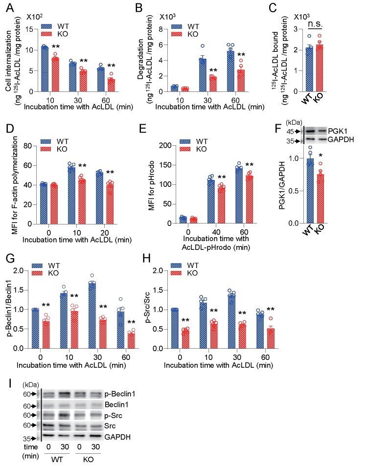
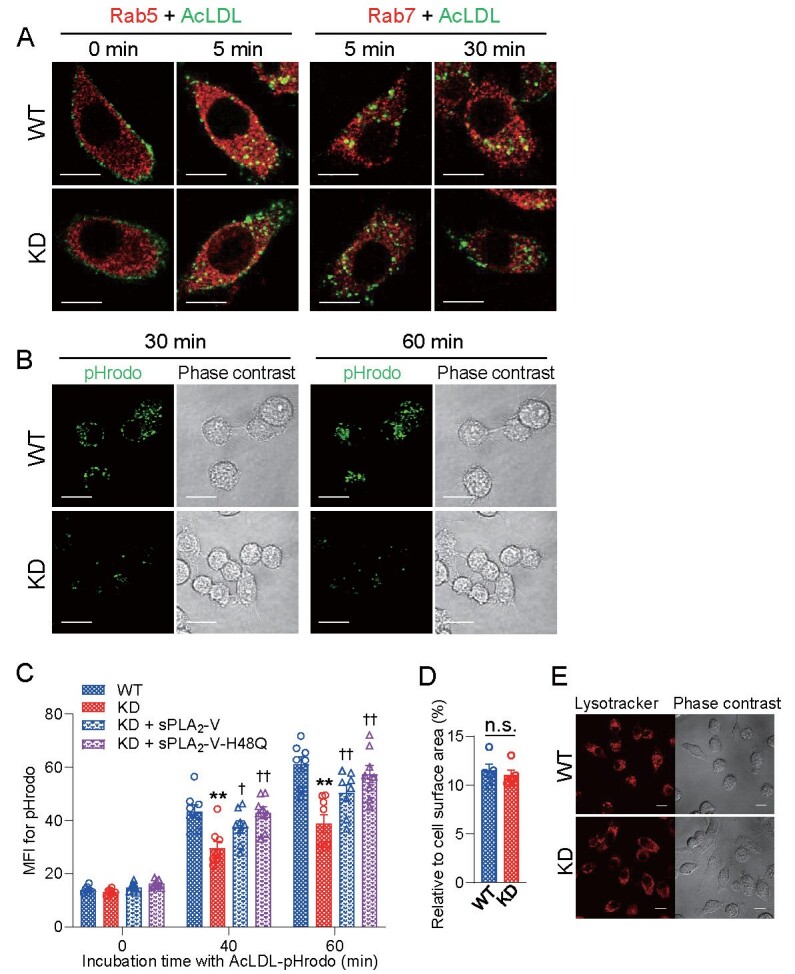
RAW264.7 cells were transfected with shRNA vector targeting sPLA-V (sPLA-V-knockdown [KD] cells) or empty vector (sPLA-V-wild-type [WT] cells). AcLDL endocytosis was assessed by incubation with I-AcLDL or AcLDL conjugated with pHrodo. Actin polymerization was assessed by flow cytometry using Alexa Fluor 546-phalloidin.
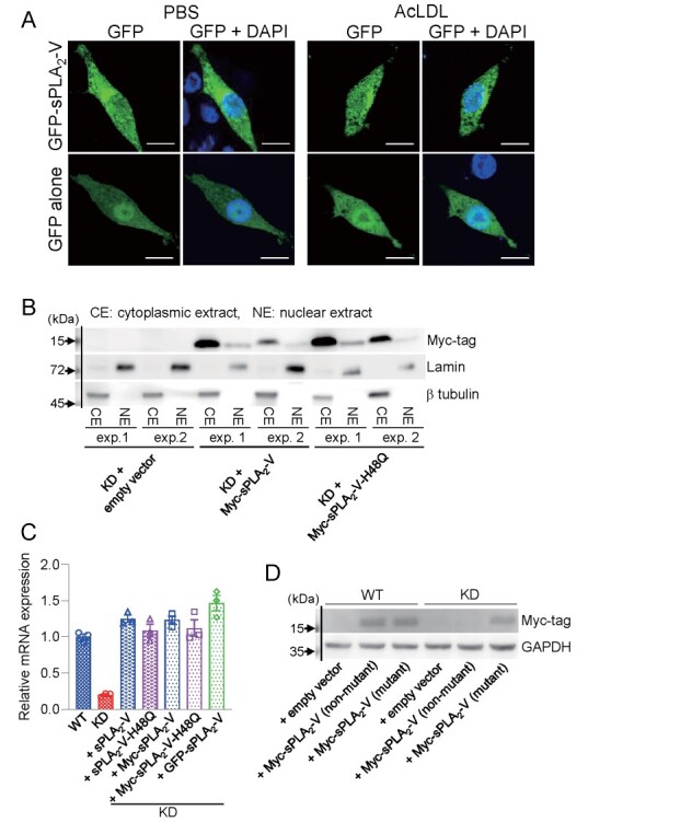
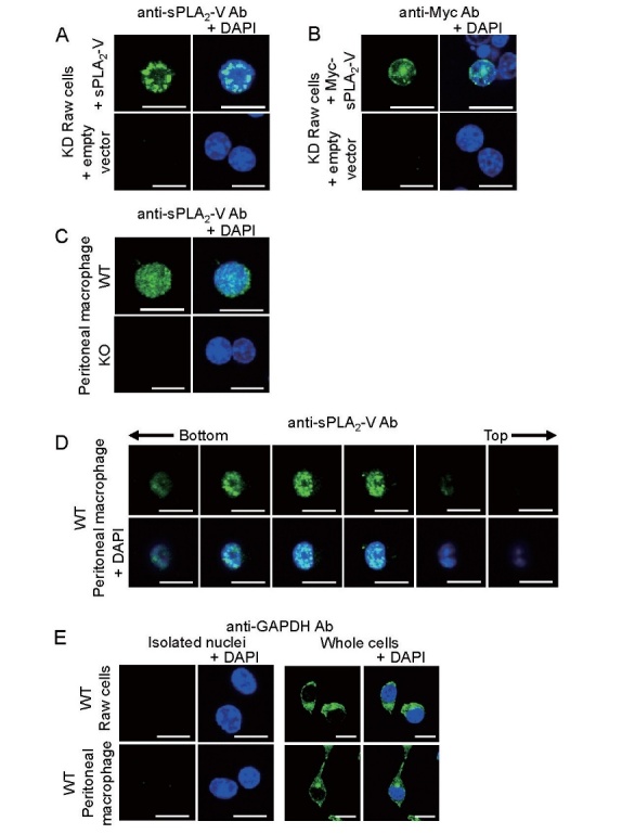
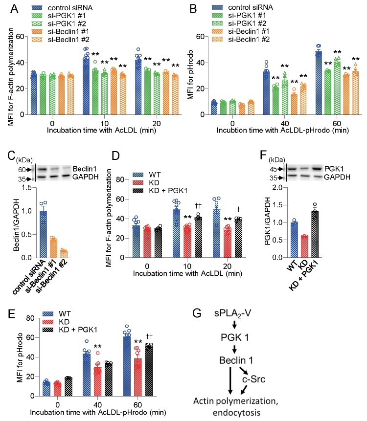
In immunofluorescence microscopic studies, sPLA-V was detected in the nucleus. ChIP-Seq and ChIP-qPCR analyses showed binding of sPLA-V to the promoter region of the phosphoglycerate kinase 1 (Pgk1) gene. In the promoter assay, sPLA-V-KD cells had lower promoter activity of the Pgk1 gene than sPLA-V-WT cells, and this decrease could be reversed by transfection with a vector encoding sPLA-V-H48Q that lacks enzymatic activity. Compared with sPLA-V-WT cells, sPLA-V-KD cells had decreased PGK1 protein expression, beclin 1 (Beclin1) phosphorylation at S30, and class III PI3-kinase activity that could also be restored by transfection with sPLA-V-H48Q. sPLA-V-KD cells had impaired actin polymerization and endocytosis, which was reversed by introduction of sPLA-V-H48Q or PGK1 overexpression. In sPLA-V-WT cells, siRNA-mediated depletion of PGK1 suppressed Beclin1 phosphorylation and impaired actin polymerization and intracellular trafficking of pHrodo-conjugated AcLDL.
Nuclear sPLA-V binds to the Pgk1 gene promoter region and increases its transcriptional activity. sPLA-V regulates AcLDL endocytosis through PGK1-Beclin1 in a manner that is independent of its enzymatic activity in RAW264.7 cells.
有人提出,第五组分泌型磷脂酶 A(sPLA-V)存在于细胞核中。本研究旨在探讨单核/巨噬细胞样细胞系 RAW264.7 细胞核 sPLA-V 是否在乙酰化低密度脂蛋白(AcLDL)的内吞作用中发挥作用。
用靶向 sPLA-V 的 shRNA 载体(sPLA-V 敲低 [KD] 细胞)或空载体(sPLA-V-野生型 [WT] 细胞)转染 RAW264.7 细胞。通过孵育与 I-AcLDL 或与 pHrodo 偶联的 AcLDL 来评估 AcLDL 的内吞作用。使用 Alexa Fluor 546-鬼笔环肽通过流式细胞术评估肌动蛋白聚合。
在免疫荧光显微镜研究中,sPLA-V 被检测到存在于细胞核中。ChIP-Seq 和 ChIP-qPCR 分析表明,sPLA-V 与磷酸甘油酸激酶 1(Pgk1)基因的启动子区域结合。在启动子分析中,与 sPLA-V-WT 细胞相比,sPLA-V-KD 细胞的 Pgk1 基因启动子活性较低,而这种降低可以通过转染缺乏酶活性的 sPLA-V-H48Q 载体来逆转。与 sPLA-V-WT 细胞相比,sPLA-V-KD 细胞的 PGK1 蛋白表达、S30 位点的 Beclin1(Beclin1)磷酸化以及 III 类 PI3-激酶活性降低,而这些可以通过转染 sPLA-V-H48Q 来恢复。sPLA-V-KD 细胞的肌动蛋白聚合和内吞作用受损,而通过引入 sPLA-V-H48Q 或 PGK1 过表达可恢复这些作用。在 sPLA-V-WT 细胞中,siRNA 介导的 PGK1 耗竭抑制了 Beclin1 磷酸化,并损害了 pHrodo 偶联的 AcLDL 的肌动蛋白聚合和细胞内转运。
细胞核 sPLA-V 与 Pgk1 基因启动子区域结合并增加其转录活性。sPLA-V 通过 PGK1-Beclin1 调节 AcLDL 的内吞作用,其方式独立于其在 RAW264.7 细胞中的酶活性。