• 文献检索
  • 文档翻译
  • 深度研究
  • 学术资讯
  • Suppr Zotero 插件Zotero 插件
  • 邀请有礼
  • 套餐&价格
  • 历史记录
应用&插件
Suppr Zotero 插件Zotero 插件浏览器插件Mac 客户端Windows 客户端微信小程序
定价
高级版会员购买积分包购买API积分包
服务
文献检索文档翻译深度研究API 文档MCP 服务
关于我们
关于 Suppr公司介绍联系我们用户协议隐私条款
关注我们

Suppr 超能文献

核心技术专利:CN118964589B侵权必究
粤ICP备2023148730 号-1Suppr @ 2026

文献检索

告别复杂PubMed语法,用中文像聊天一样搜索,搜遍4000万医学文献。AI智能推荐,让科研检索更轻松。

立即免费搜索

文件翻译

保留排版,准确专业,支持PDF/Word/PPT等文件格式,支持 12+语言互译。

免费翻译文档

深度研究

AI帮你快速写综述,25分钟生成高质量综述,智能提取关键信息,辅助科研写作。

立即免费体验

疑似特发性肺纤维化患者的实用影像解读:肺纤维化基金会放射学工作组的官方建议

Practical Imaging Interpretation in Patients Suspected of Having Idiopathic Pulmonary Fibrosis: Official Recommendations from the Radiology Working Group of the Pulmonary Fibrosis Foundation.

作者信息

Hobbs Stephen, Chung Jonathan H, Leb Jay, Kaproth-Joslin Kate, Lynch David A

机构信息

Department of Radiology, University of Kentucky, 800 Rose St, HX-315B, Lexington, KY 40536 (S.H.); Department of Radiology, University of Chicago, Chicago, Ill (J.H.C.); Department of Radiology, Columbia University, New York, NY (J.L.); Department of Imaging Sciences, University of Rochester, Rochester, NY (K.K.J.); and Department of Radiology, National Jewish Health, Denver, Colo (D.A.L.).

出版信息

Radiol Cardiothorac Imaging. 2021 Feb 25;3(1):e200279. doi: 10.1148/ryct.2021200279. eCollection 2021 Feb.

DOI:10.1148/ryct.2021200279
PMID:33778653
原文链接:https://pmc.ncbi.nlm.nih.gov/articles/PMC7977697/
Abstract

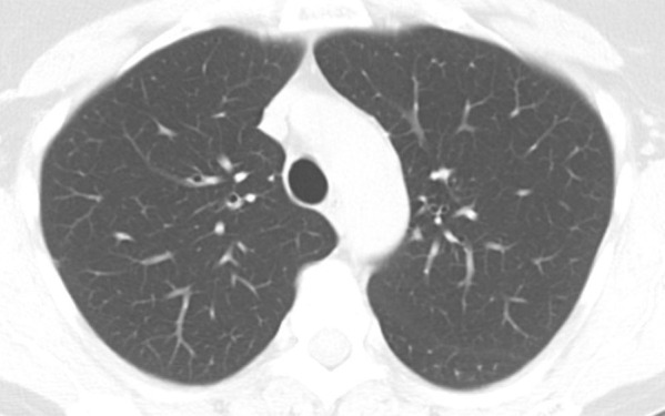
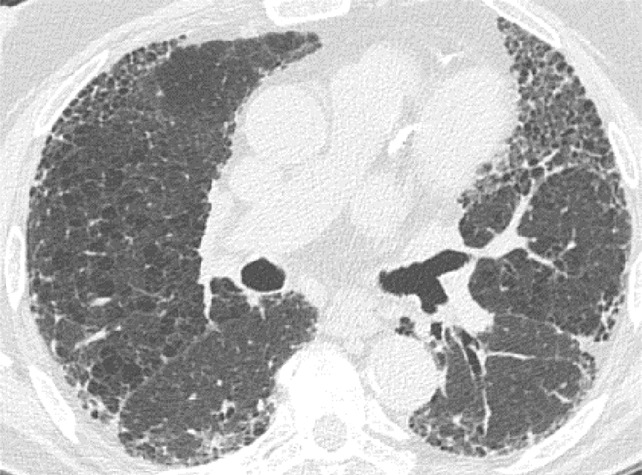
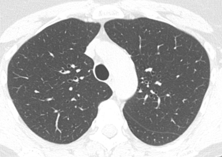
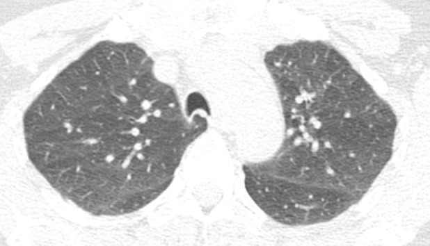
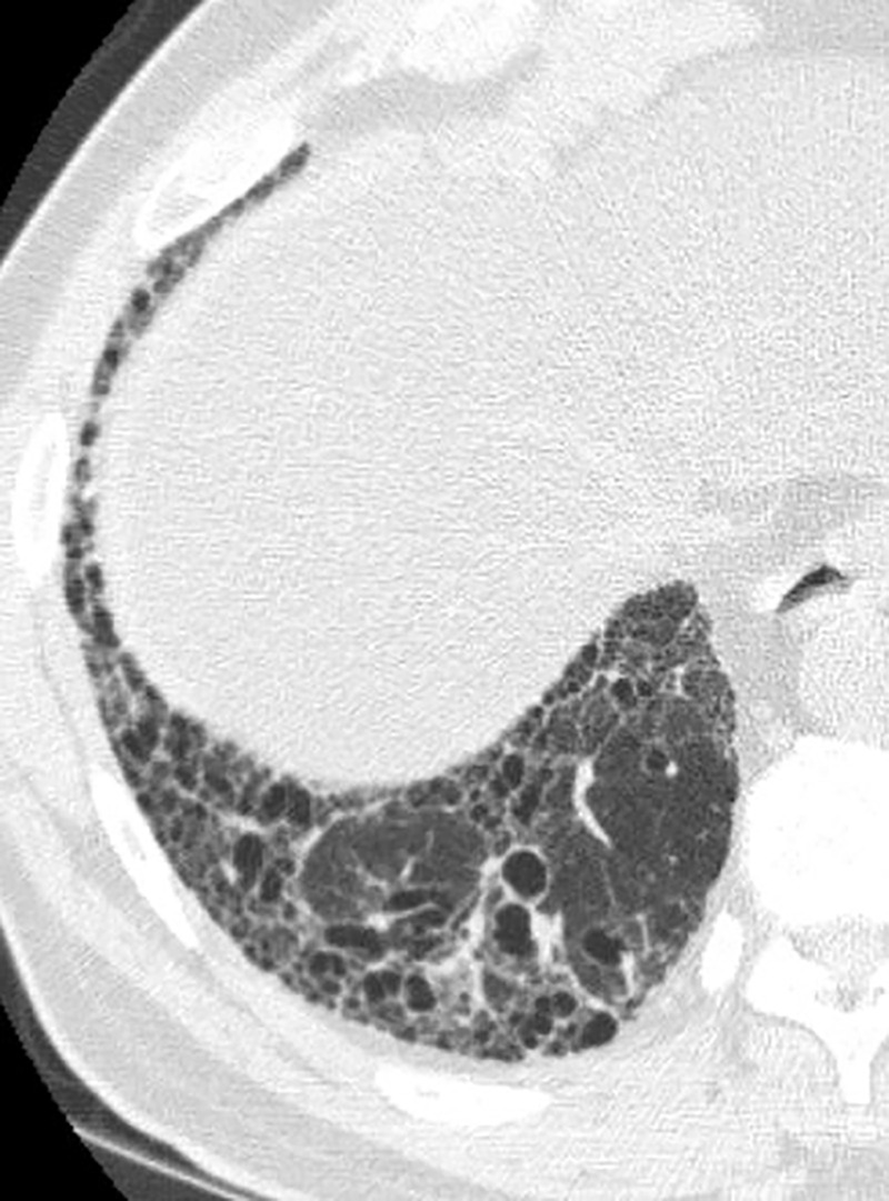
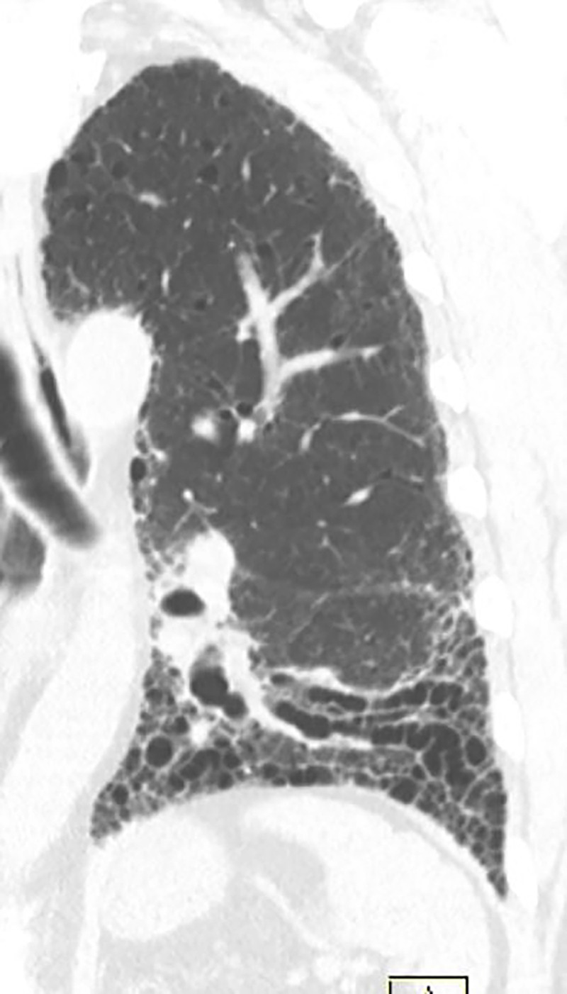
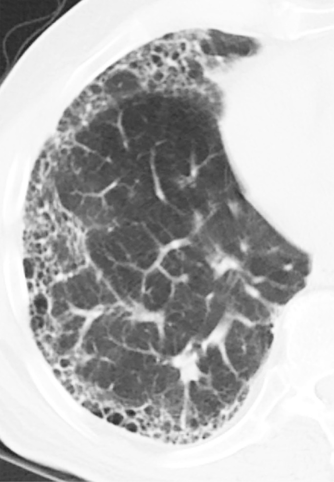
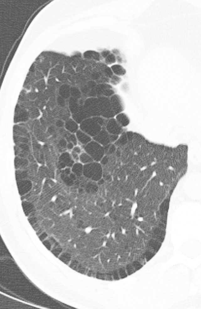
Imaging serves a key role in the diagnosis of patients suspected of having idiopathic pulmonary fibrosis (IPF). Accurate pattern classification at thin-section chest CT is a key step in multidisciplinary discussions, guiding the need for surgical lung biopsy and determining available pharmacologic therapies. The recent approval of new treatments for fibrosing lung disease has made it more critical than ever for radiologists to facilitate accurate and early diagnosis of IPF. This document was developed by the Radiology Working Group of the Pulmonary Fibrosis Foundation with the goal of providing a practical guide for radiologists. In this review, the critical imaging patterns of IPF, pitfalls in imaging classifications, confounding imaging findings with other fibrotic lung diseases, and reporting standards for cases of lung fibrosis will be discussed. Published under a CC BY 4.0 license. See also the commentary by White and Galvin in this issue.

摘要

影像学在疑似特发性肺纤维化(IPF)患者的诊断中起着关键作用。薄层胸部CT上准确的模式分类是多学科讨论的关键步骤,指导手术肺活检的必要性并确定可用的药物治疗方法。最近用于纤维化性肺病的新疗法获批,使得放射科医生比以往任何时候都更迫切需要促进IPF的准确和早期诊断。本文档由肺纤维化基金会放射学工作组编写,目的是为放射科医生提供实用指南。在本综述中,将讨论IPF的关键影像学模式、影像学分型中的陷阱、与其他纤维化性肺病混淆的影像学表现以及肺纤维化病例的报告标准。根据知识共享署名4.0许可协议发布。另见本期怀特和加尔文的评论。

https://cdn.ncbi.nlm.nih.gov/pmc/blobs/374f/7977697/d2db49beff84/ryct.2021200279.fig9c.jpg
https://cdn.ncbi.nlm.nih.gov/pmc/blobs/374f/7977697/7146fbb431b4/ryct.2021200279.fig1a.jpg
https://cdn.ncbi.nlm.nih.gov/pmc/blobs/374f/7977697/b49e855d1caf/ryct.2021200279.fig1b.jpg
https://cdn.ncbi.nlm.nih.gov/pmc/blobs/374f/7977697/8b1031b8d98f/ryct.2021200279.fig1c.jpg
https://cdn.ncbi.nlm.nih.gov/pmc/blobs/374f/7977697/252e80f116c9/ryct.2021200279.fig2.jpg
https://cdn.ncbi.nlm.nih.gov/pmc/blobs/374f/7977697/9b65c9756b3b/ryct.2021200279.fig3.jpg
https://cdn.ncbi.nlm.nih.gov/pmc/blobs/374f/7977697/00c73edff7af/ryct.2021200279.fig4a.jpg
https://cdn.ncbi.nlm.nih.gov/pmc/blobs/374f/7977697/6d755a7141f3/ryct.2021200279.fig4b.jpg
https://cdn.ncbi.nlm.nih.gov/pmc/blobs/374f/7977697/55922be7b2aa/ryct.2021200279.fig5.jpg
https://cdn.ncbi.nlm.nih.gov/pmc/blobs/374f/7977697/556068393f13/ryct.2021200279.fig6.jpg
https://cdn.ncbi.nlm.nih.gov/pmc/blobs/374f/7977697/46ea06580f43/ryct.2021200279.fig7a.jpg
https://cdn.ncbi.nlm.nih.gov/pmc/blobs/374f/7977697/6f5e8a6bb8d7/ryct.2021200279.fig7b.jpg
https://cdn.ncbi.nlm.nih.gov/pmc/blobs/374f/7977697/1dfd0f1c883a/ryct.2021200279.fig7c.jpg
https://cdn.ncbi.nlm.nih.gov/pmc/blobs/374f/7977697/91505484d440/ryct.2021200279.fig8a.jpg
https://cdn.ncbi.nlm.nih.gov/pmc/blobs/374f/7977697/39d1b949ef2c/ryct.2021200279.fig8b.jpg
https://cdn.ncbi.nlm.nih.gov/pmc/blobs/374f/7977697/a7723ec6b166/ryct.2021200279.fig9a.jpg
https://cdn.ncbi.nlm.nih.gov/pmc/blobs/374f/7977697/ac7cca2f81f5/ryct.2021200279.fig9b.jpg
https://cdn.ncbi.nlm.nih.gov/pmc/blobs/374f/7977697/d2db49beff84/ryct.2021200279.fig9c.jpg

相似文献

1
Practical Imaging Interpretation in Patients Suspected of Having Idiopathic Pulmonary Fibrosis: Official Recommendations from the Radiology Working Group of the Pulmonary Fibrosis Foundation.疑似特发性肺纤维化患者的实用影像解读:肺纤维化基金会放射学工作组的官方建议
Radiol Cardiothorac Imaging. 2021 Feb 25;3(1):e200279. doi: 10.1148/ryct.2021200279. eCollection 2021 Feb.
2
Idiopathic pulmonary fibrosis: the radiologist's role in making the diagnosis.特发性肺纤维化:放射科医生在诊断中的作用。
Br J Radiol. 2019 Jul;92(1099):20181003. doi: 10.1259/bjr.20181003. Epub 2019 May 14.
3
Diagnostic certainty of idiopathic pulmonary fibrosis/usual interstitial pneumonia: The effect of the integrated clinico-radiological assessment.特发性肺纤维化/普通间质性肺炎的诊断确定性:综合临床-放射学评估的影响
Eur J Radiol. 2015 Dec;84(12):2640-5. doi: 10.1016/j.ejrad.2015.08.016. Epub 2015 Sep 1.
4
Chronic hypersensitivity pneumonitis in patients diagnosed with idiopathic pulmonary fibrosis: a prospective case-cohort study.特发性肺纤维化患者的慢性过敏性肺炎:一项前瞻性病例队列研究。
Lancet Respir Med. 2013 Nov;1(9):685-94. doi: 10.1016/S2213-2600(13)70191-7. Epub 2013 Oct 21.
5
Bronchoscopic Lung Cryobiopsy Increases Diagnostic Confidence in the Multidisciplinary Diagnosis of Idiopathic Pulmonary Fibrosis.支气管镜肺冷冻活检增加特发性肺纤维化多学科诊断的信心。
Am J Respir Crit Care Med. 2016 Apr 1;193(7):745-52. doi: 10.1164/rccm.201504-0711OC.
6
Diagnostic criteria for idiopathic pulmonary fibrosis: a Fleischner Society White Paper.特发性肺纤维化的诊断标准:弗利彻协会白皮书。
Lancet Respir Med. 2018 Feb;6(2):138-153. doi: 10.1016/S2213-2600(17)30433-2. Epub 2017 Nov 15.
7
Predictors of idiopathic pulmonary fibrosis in absence of radiologic honeycombing: A cross sectional analysis in ILD patients undergoing lung tissue sampling.无放射学蜂窝状改变的特发性肺纤维化的预测因素:对接受肺组织采样的间质性肺疾病患者的横断面分析
Respir Med. 2016 Sep;118:88-95. doi: 10.1016/j.rmed.2016.07.016. Epub 2016 Jul 28.
8
Idiopathic pulmonary fibrosis: current and future directions.特发性肺纤维化:现状与未来方向
Clin Radiol. 2017 May;72(5):343-355. doi: 10.1016/j.crad.2016.12.014. Epub 2017 Jan 21.
9
Deep learning for classifying fibrotic lung disease on high-resolution computed tomography: a case-cohort study.深度学习在高分辨率 CT 上对肺纤维化疾病的分类:一项病例队列研究。
Lancet Respir Med. 2018 Nov;6(11):837-845. doi: 10.1016/S2213-2600(18)30286-8. Epub 2018 Sep 16.
10
Multicentre evaluation of multidisciplinary team meeting agreement on diagnosis in diffuse parenchymal lung disease: a case-cohort study.多中心评价弥漫性实质性肺疾病多学科团队会议诊断协议:病例队列研究。
Lancet Respir Med. 2016 Jul;4(7):557-565. doi: 10.1016/S2213-2600(16)30033-9. Epub 2016 May 11.

引用本文的文献

1
Classifying chronic obstructive pulmonary disease status using computed tomography imaging and convolutional neural networks: comparison of model input image types and training data severity.使用计算机断层扫描成像和卷积神经网络对慢性阻塞性肺疾病状态进行分类:模型输入图像类型与训练数据严重程度的比较
J Med Imaging (Bellingham). 2025 May;12(3):034502. doi: 10.1117/1.JMI.12.3.034502. Epub 2025 May 22.
2
Traditional Herbal Plants and their Phytoconstituents Based Remedies for Respiratory Diseases: A Review.基于传统草药植物及其植物成分的呼吸系统疾病治疗方法综述
Open Respir Med J. 2025 Feb 12;19:e18743064341009. doi: 10.2174/0118743064341009241210045737. eCollection 2025.
3

本文引用的文献

1
Magnetic resonance imaging of interstitial lung diseases: A state-of-the-art review.间质性肺疾病的磁共振成像:最新综述。
Respir Med. 2019 Aug;155:79-85. doi: 10.1016/j.rmed.2019.07.006. Epub 2019 Jul 6.
2
Idiopathic pulmonary fibrosis: the radiologist's role in making the diagnosis.特发性肺纤维化:放射科医生在诊断中的作用。
Br J Radiol. 2019 Jul;92(1099):20181003. doi: 10.1259/bjr.20181003. Epub 2019 May 14.
3
New Developments in Imaging Idiopathic Pulmonary Fibrosis With Hyperpolarized Xenon Magnetic Resonance Imaging.
Anti-neutrophil cytoplasmic antibodies associated interstitial pneumonia: A possible new clinical entity.
抗中性粒细胞胞浆抗体相关性间质性肺炎:一种可能的新临床实体。
Eur J Clin Invest. 2025 Jul;55(7):e70025. doi: 10.1111/eci.70025. Epub 2025 Mar 20.
4
Pixel-Wise Interstitial Lung Disease Interval Change Analysis: A Quantitative Evaluation Method for Chest Radiographs Using Weakly Supervised Learning.逐像素间质性肺疾病间隔变化分析:一种使用弱监督学习的胸部X光片定量评估方法。
Bioengineering (Basel). 2024 Jun 2;11(6):562. doi: 10.3390/bioengineering11060562.
5
Imaging in the diagnosis and management of fibrosing interstitial lung diseases.纤维化间质性肺疾病诊断与管理中的影像学检查
Breathe (Sheff). 2024 Mar;20(1):240006. doi: 10.1183/20734735.0006-2024. Epub 2024 May 14.
6
Thin-Section CT in the Categorization and Management of Pulmonary Fibrosis including Recently Defined Progressive Pulmonary Fibrosis.薄层 CT 在肺纤维化的分类和管理中的应用,包括最近定义的进行性肺纤维化。
Radiol Cardiothorac Imaging. 2024 Feb;6(1):e230135. doi: 10.1148/ryct.230135.
7
Developing the Lung Graph-Based Machine Learning Model for Identification of Fibrotic Interstitial Lung Diseases.开发基于肺图的机器学习模型以识别纤维性间质性肺疾病。
J Imaging Inform Med. 2024 Feb;37(1):268-279. doi: 10.1007/s10278-023-00909-7. Epub 2024 Jan 16.
8
Improving Prognostication in Pulmonary Hypertension Using AI-quantified Fibrosis and Radiologic Severity Scoring at Baseline CT.利用 AI 量化的纤维化和基线 CT 影像学严重程度评分改善肺动脉高压的预后。
Radiology. 2024 Feb;310(2):e231718. doi: 10.1148/radiol.231718.
9
Delayed inflammatory pulmonary syndrome: A distinct clinical entity in the spectrum of inflammatory syndromes in COVID-19 infection?迟发性炎症性肺综合征:新型冠状病毒肺炎感染炎症综合征谱中的一种独特临床实体?
World J Crit Care Med. 2023 Sep 9;12(4):226-235. doi: 10.5492/wjccm.v12.i4.226.
10
Deep Learning Assessment of Progression of Emphysema and Fibrotic Interstitial Lung Abnormality.深度学习评估肺气肿和纤维性间质肺异常的进展。
Am J Respir Crit Care Med. 2023 Sep 15;208(6):666-675. doi: 10.1164/rccm.202211-2098OC.
用超极化氙磁共振成像技术对特发性肺纤维化的新进展成像。
J Thorac Imaging. 2019 Mar;34(2):136-150. doi: 10.1097/RTI.0000000000000392.
4
Imaging in idiopathic pulmonary fibrosis: diagnosis and mimics.特发性肺纤维化的影像学表现:诊断与鉴别诊断。
Clinics (Sao Paulo). 2019 Feb 4;74:e225. doi: 10.6061/clinics/2019/e225.
5
Diagnosis of Idiopathic Pulmonary Fibrosis. An Official ATS/ERS/JRS/ALAT Clinical Practice Guideline.特发性肺纤维化诊断。美国胸科学会/欧洲呼吸学会/日本呼吸学会/拉丁美洲胸科学会临床实践指南。
Am J Respir Crit Care Med. 2018 Sep 1;198(5):e44-e68. doi: 10.1164/rccm.201807-1255ST.
6
Diagnostic criteria for idiopathic pulmonary fibrosis: a Fleischner Society White Paper.特发性肺纤维化的诊断标准:弗利彻协会白皮书。
Lancet Respir Med. 2018 Feb;6(2):138-153. doi: 10.1016/S2213-2600(17)30433-2. Epub 2017 Nov 15.
7
Effects of pure and hybrid iterative reconstruction algorithms on high-resolution computed tomography in the evaluation of interstitial lung disease.纯迭代重建算法和混合迭代重建算法在间质性肺疾病高分辨率计算机断层扫描评估中的作用。
Eur J Radiol. 2017 Aug;93:243-251. doi: 10.1016/j.ejrad.2017.06.003. Epub 2017 Jun 4.
8
Idiopathic Pulmonary Fibrosis: Data-driven Textural Analysis of Extent of Fibrosis at Baseline and 15-Month Follow-up.特发性肺纤维化:基于数据的基线和15个月随访时纤维化范围的纹理分析
Radiology. 2017 Oct;285(1):270-278. doi: 10.1148/radiol.2017161177. Epub 2017 May 10.
9
Familial pulmonary fibrosis: Clinical and radiological characteristics and progression analysis in different high resolution-CT patterns.家族性肺纤维化:不同高分辨率CT模式下的临床和放射学特征及进展分析
Respir Med. 2017 May;126:75-83. doi: 10.1016/j.rmed.2017.03.020. Epub 2017 Mar 22.
10
The use of pretest probability increases the value of high-resolution CT in diagnosing usual interstitial pneumonia.使用检验前概率可提高高分辨率CT在诊断普通型间质性肺炎中的价值。
Thorax. 2017 May;72(5):424-429. doi: 10.1136/thoraxjnl-2016-209671. Epub 2017 Jan 12.