University of Zurich, Zurich, Switzerland.
Center for MR-Research, University Children's Hospital, Steinwiesstrasse 75, 8032, Zurich, Switzerland.
Eur Radiol. 2021 Oct;31(10):7231-7241. doi: 10.1007/s00330-021-07813-0. Epub 2021 Mar 30.
To use 4D-flow MRI to describe systemic and non-systemic ventricular flow organisation and energy loss in patients with repaired d-transposition of the great arteries (d-TGA) and normal subjects.
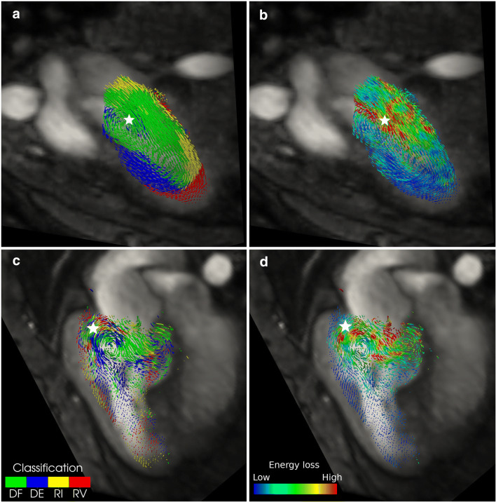
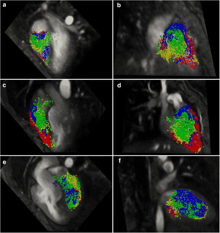
Pathline tracking of ventricular volumes was performed using 4D-flow MRI data from a 1.5-T GE Discovery MR450 scanner. D-TGA patients following arterial switch (n = 17, mean age 14 ± 5 years) and atrial switch (n = 15, 35 ± 6 years) procedures were examined and compared with subjects with normal cardiac anatomy and ventricular function (n = 12, 12 ± 3 years). Pathlines were classified by their passage through the ventricles as direct flow, retained inflow, delayed ejection flow, and residual volume and visually and quantitatively assessed. Additionally, viscous energy losses (EL) were calculated.
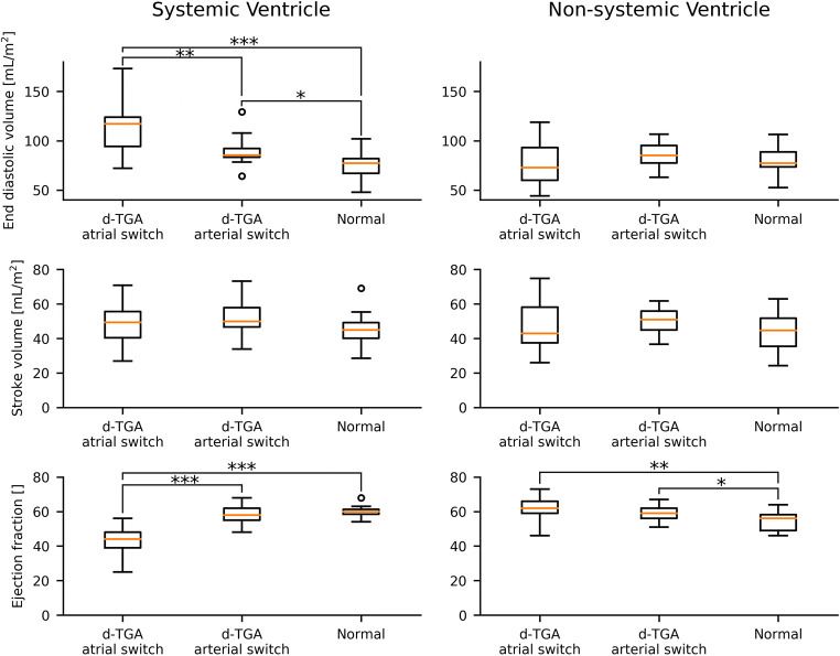
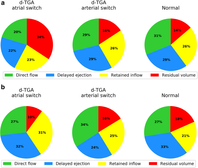
In normal subjects, the ventricular flow paths were well ordered following similar trajectories through the ventricles with very little mixing of flow components. The flow paths in all atrial and some arterial switch patients were more irregular with high mixing. Direct flow and delayed ejection flow were decreased in atrial switch patients' systemic ventricles with a corresponding increase in residual volume compared with normal subjects (p = 0.003 and p < 0.001 respectively) and arterial switch patients (p < 0.0001 and p < 0.001 respectively). In non-systemic ventricles, arterial switch patients had increased direct flow and decreased delayed ejection fractions compared to normal (p = 0.007 and p < 0.001 respectively) and atrial switch patients (p = 0.01 and p < 0.001 respectively). Regions of high levels of mixing of ventricular flow components showed elevated EL.
4D-flow MRI pathline tracking reveals disordered ventricular flow patterns and associated EL in d-TGA patients.
• 4D-flow MRI can be used to assess intraventricular flow dynamics in d-TGA patients. • d-TGA arterial switch patients mostly show intraventricular flow dynamics representative of normal subjects, while atrial switch patients show increased flow disorder and different proportions of intraventricular flow volumes. • Flow disruption and disorder increase viscous energy losses.
使用 4D-flow MRI 描述修复后的右旋位大动脉转位(d-TGA)患者和正常受试者的系统性和非系统性心室流动组织和能量损失。
使用 1.5-T GE Discovery MR450 扫描仪的 4D-flow MRI 数据对心室容积进行路径线追踪。检查了动脉切换(n = 17,平均年龄 14 ± 5 岁)和心房切换(n = 15,35 ± 6 岁)手术后的 d-TGA 患者,并与具有正常心脏解剖结构和心室功能的受试者(n = 12,12 ± 3 岁)进行了比较。根据路径线穿过心室的情况,将路径线分为直接流动、滞留流入、延迟射流和残余容积,并进行了视觉和定量评估。此外,还计算了粘性能量损失(EL)。
在正常受试者中,心室流动路径通过心室的相似轨迹有序排列,流动成分的混合很少。所有心房和一些动脉切换患者的流动路径更加不规则,混合度更高。与正常受试者(p = 0.003 和 p < 0.001)和动脉切换患者(p < 0.0001 和 p < 0.001)相比,心房切换患者的系统性心室中的直接流动和延迟射流减少,而残余容积增加(p = 0.003 和 p < 0.001)。在非系统性心室中,与正常(p = 0.007 和 p < 0.001)和心房切换(p = 0.01 和 p < 0.001)患者相比,动脉切换患者的直接流动增加,而延迟射流分数减少。显示心室流动成分混合程度较高的区域显示出较高的 EL。
4D-flow MRI 路径线追踪显示 d-TGA 患者的心室流动模式紊乱和相关的 EL。
• 4D-flow MRI 可用于评估 d-TGA 患者的心室内流动动力学。• d-TGA 动脉切换患者的心室内流动动力学主要表现为正常受试者的代表,而心房切换患者则显示出更高的流动紊乱和不同比例的心室内流动量。• 流动中断和紊乱会增加粘性能量损失。