Department of Ultrasound.
Department of Pathology, West China Hospital of Sichuan University, No. 37 Guo Xue Xiang, Chengdu, Sichuan Province, China.
Medicine (Baltimore). 2021 Apr 2;100(13):e25178. doi: 10.1097/MD.0000000000025178.
Fungal liver infection mostly occurs in immunocompromised patients, and is often associated with delayed diagnosis and high mortality rates. Dynamic contrast enhanced imaging is crucial for the diagnosis of fungal liver infection and has been reported having variable manifestations.
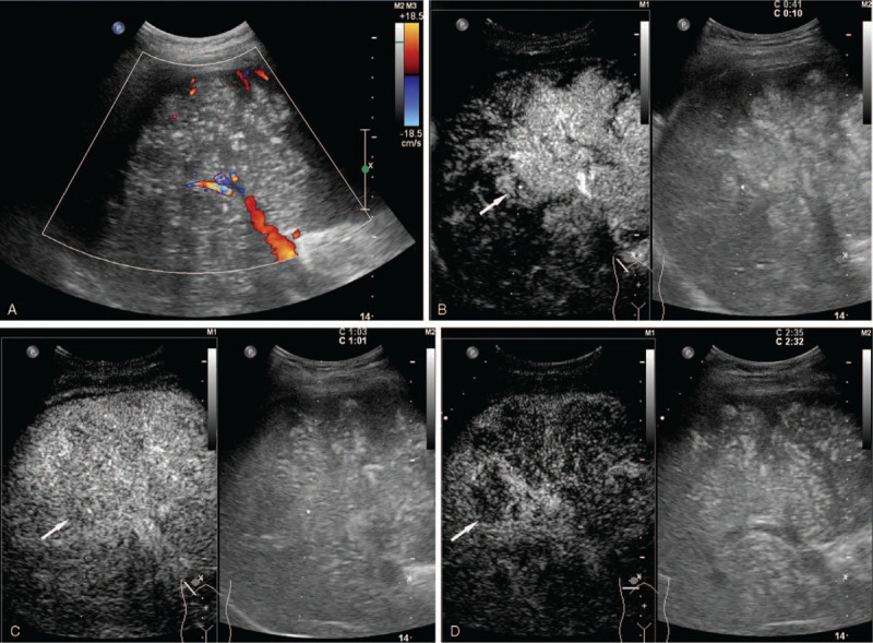
A 38-year-old Chinese man, with a history of diabetes and chronic hepatitis B, was admitted to our hospital due to prolonged fever of unknown cause. He had a medical history of receiving broad-spectrum antibiotic treatment for pulmonary inflammation at the local hospital. The blood test results showed that the white cell count (14.0 × 10⁹/L) and neutrophil count ratio (77.0%) were subtly elevated. C-reactive protein (92.0 mg/l) and cancer antigen (CA)-125 (904.50 U/ml) were elevated. Non-small cell lung cancer antigen was within the normal limit. Hepatitis B virus DNA load was 3.28 × 10³ IU/ml. Sputum and blood cultures were normal. Abdominal ultrasonography (US) found a large heterogeneous mass, with diffused echogenic foci without infiltrating the surrounding vascular, which exhibiting "rapid wash in and out" on contrast-enhanced ultrasound (CEUS).
The diagnosis of liver fungal infection was confirmed pathologically via ultrasound-guided biopsy.
Antibiotic and antifungal therapy with imipenem and voriconazole.
The patient's body temperature had been controlled and the huge mass disappeared on follow-up ultrasound 1-year later.
This case highlights the unusual imaging features of fungal liver infection, presenting as huge heterogeneous mass with diffusive echogenic foci without infiltrating the surrounding vascular on grayscale US and the enhancement pattern of "rapid wash in and out" on CEUS. Additionally, ultrasound-guided biopsy is necessary for the correct diagnosis of suspected liver lesions.
真菌感染性肝脏疾病主要发生于免疫功能低下的患者,常因诊断延迟而导致病死率较高。动态对比增强成像对于真菌感染性肝脏疾病的诊断具有重要价值,其影像学表现具有多样性。
一名 38 岁中国男性,患有糖尿病和慢性乙型肝炎,因不明原因的长期发热于我院就诊。他曾在当地医院因肺部炎症接受过广谱抗生素治疗。血液检查结果显示,白细胞计数(14.0×10⁹/L)和中性粒细胞比例(77.0%)略有升高。C 反应蛋白(92.0mg/l)和癌抗原 125(904.50U/ml)升高。非小细胞肺癌抗原在正常范围内。乙型肝炎病毒 DNA 载量为 3.28×10³IU/ml。痰和血培养均正常。腹部超声检查发现巨大的混杂回声肿块,弥漫性回声增强灶,无周围血管浸润,超声造影表现为“快进快出”。
经超声引导活检病理确诊为肝脏真菌感染。
给予亚胺培南和伏立康唑进行抗生素和抗真菌治疗。
患者体温得到控制,1 年后随访超声发现巨大肿块消失。
本病例提示真菌感染性肝脏疾病的影像学表现较为特殊,表现为灰阶超声上巨大的混杂回声肿块,弥漫性回声增强灶,无周围血管浸润,超声造影表现为“快进快出”。此外,超声引导活检对于疑似肝脏病变的正确诊断是必要的。