Department of Nuclear Medicine, Charité-Universitätsmedizin Berlin, Berlin, Germany.
Department of Radiation Oncology, Charité-Universitätsmedizin Berlin, Berlin, Germany.
F1000Res. 2020 Nov 19;9:1350. doi: 10.12688/f1000research.27303.2. eCollection 2020.
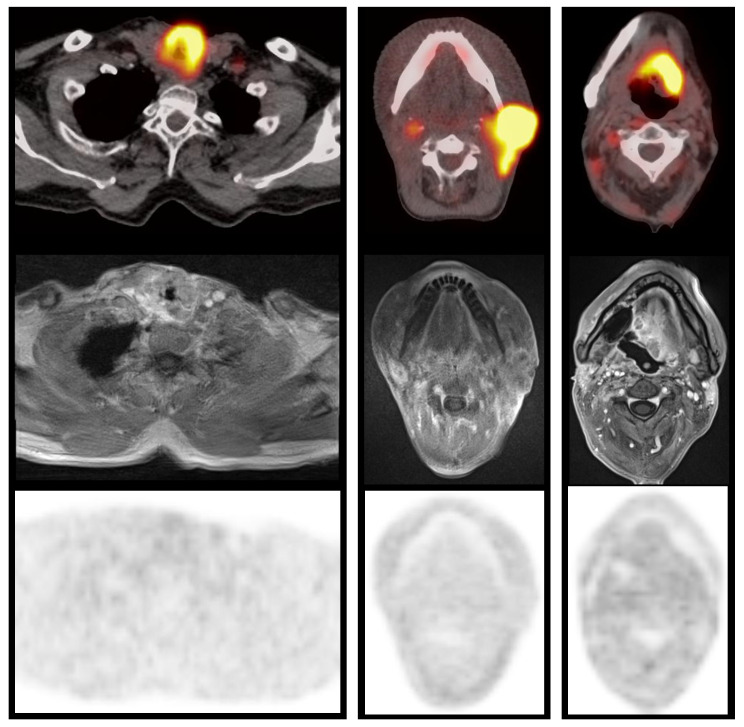
Tumor hypoxia measured by dedicated tracers like [ F]fluoromisonidazole (FMISO) is a well-established prognostic factor in head and neck squamous cell carcinomas (HNSCC) treated with definitive chemoradiation (CRT). However, prevalence and characteristics of positron emission tomography (PET) measured hypoxia in patients with relapse after previous irradiation is missing. Here we report imaging findings of a prospective pilot study in HNSCC patients treated with re-irradiation. In 8 patients with recurrent HNSCC, diagnosed at a median of 18 months after initial radiotherapy/CRT, [ F]fluorodeoxyglucose (FDG)-PET/CT (n=8) and FMISO-PET/MRI (n=7) or FMISO-PET/CT (n=1) were performed. Static FMISO-PET was performed after 180 min. MRI sequences in PET/MRI included diffusion-weighted imaging with apparent diffusion coefficient (ADC) values and contrast enhanced T1w imaging (StarVIBE). Lesions (primary tumor recurrence, 4; cervical lymph node, 1; both, 3) were delineated on FDG-PET and FMISO-PET data using a background-adapted threshold-based method. SUV and SUV in FDG- and FMISO-PET were derived, as well as maximum tumor-to-muscle ratio (TMR ) and hypoxic volume with 1.6-fold muscle SUV (HV ) in FMISO-PET. Intensity of lesional contrast enhancement was rated relative to contralateral normal tissue. Average ADC values were derived from a 2D region of interest in the tumor. In FMISO-PET, median TMR was 1.7 (range: 1.1-1.8). Median HV was 0.05 ml (range: 0-7.3 ml). Only in 2/8 patients, HV was ≥1.0 ml. In FDG-PET, median SUV was 9.3 (range: 5.0-20.1). On contrast enhanced imaging four lesions showed decreased and four lesions increased contrast enhancement compared to non-pathologic reference tissue. Median average ADC was 1,060 ×10 mm /s (range: 840-1,400 ×10 mm /s). This pilot study implies that hypoxia detectable by FMISO-PET may not be as prevalent as expected among loco-regional recurrent, HPV negative HNSCC. ADC values were only mildly reduced, and contrast enhancement was variable. The results require confirmation in larger sample sizes.
肿瘤乏氧程度可通过专用示踪剂进行测量,如[ F]氟代米索硝唑(FMISO),这是头颈部鳞状细胞癌(HNSCC)接受根治性放化疗(CRT)后一种成熟的预后因素。然而,在先前接受过放疗的患者中,复发后 PET 测量的乏氧程度的发生率和特征尚不清楚。本研究报道了一项在接受再放疗的 HNSCC 患者中进行的前瞻性试点研究的影像学结果。8 例局部复发的 HNSCC 患者(中位时间为初始放疗/CRT 后 18 个月),进行了[ F]氟脱氧葡萄糖(FDG)-PET/CT(n=8)和 FMISO-PET/MRI(n=7)或 FMISO-PET/CT(n=1)。在 180 分钟后进行 FMISO 静态 PET。PET/MRI 的 MRI 序列包括扩散加权成像(DWI)和表观扩散系数(ADC)值,以及对比增强 T1w 成像(StarVIBE)。在 FDG-PET 和 FMISO-PET 数据上使用基于背景适应的阈值方法对病灶(原发肿瘤复发 4 例,颈部淋巴结转移 1 例,两者均有 3 例)进行了勾画。FDG-PET 和 FMISO-PET 中均推导了 SUV 和 SUV,以及 FMISO-PET 中 1.6 倍肌肉 SUV(HV)的最大肿瘤与肌肉比(TMR)和乏氧体积。病变强化的强度与对侧正常组织进行了比较。肿瘤的二维 ROI 中获得了平均 ADC 值。FMISO-PET 中,TMR 的中位数为 1.7(范围:1.1-1.8)。HV 的中位数为 0.05ml(范围:0-7.3ml)。只有 2/8 例患者的 HV 大于 1.0ml。FDG-PET 中,SUV 的中位数为 9.3(范围:5.0-20.1)。在对比增强成像中,与非病理性参考组织相比,4 个病灶的强化程度降低,4 个病灶的强化程度增加。平均 ADC 的中位数为 1060×10 mm /s(范围:840-1400×10 mm /s)。这项试点研究表明,在局部复发、HPV 阴性的 HNSCC 患者中,FMISO-PET 检测到的乏氧可能并不像预期的那样普遍。ADC 值仅略有降低,强化程度也存在差异。这些结果需要在更大的样本量中进行验证。