Department of Radiation Oncology, Medical Center University of Freiburg, Faculty of Medicine, University of Freiburg, Freiburg, Germany.
German Cancer Consortium (DKTK), Partner Site Freiburg, Freiburg, Germany.
Radiat Oncol. 2018 Aug 29;13(1):159. doi: 10.1186/s13014-018-1103-1.
To assess the effect of radiochemotherapy (RCT) on proposed tumour hypoxia marker transverse relaxation time (T2*) and to analyse the relation between T2* and F-misonidazole PET/CT (FMISO-PET) and F-fluorodeoxyglucose PET/CT (FDG-PET).
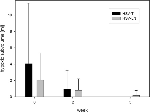
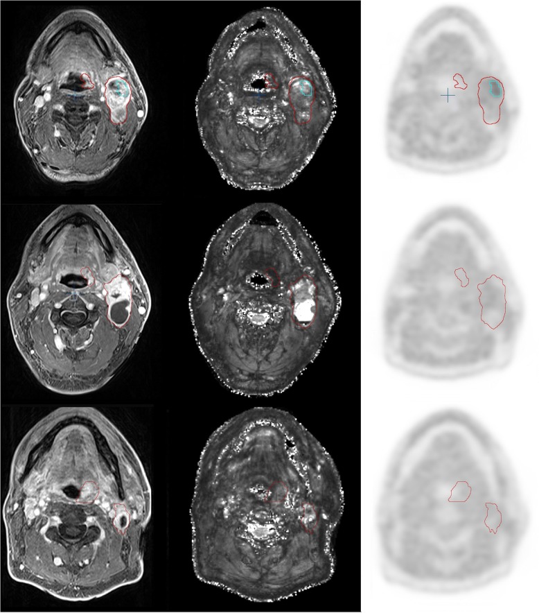
Ten patients undergoing definitive RCT for squamous cell head-and-neck cancer (HNSCC) received repeat FMISO- and 3 Tesla T2*-weighted MRI at weeks 0, 2 and 5 during treatment and FDG-PET at baseline. Gross tumour volumes (GTV) of tumour (T), lymph nodes (LN) and hypoxic subvolumes (HSV, based on FMISO-PET) and complementary non-hypoxic subvolumes (nonHSV) were generated. Mean values for T2* and SUVmean FDG were determined.
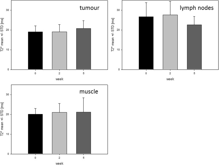
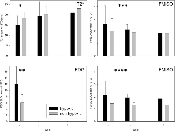
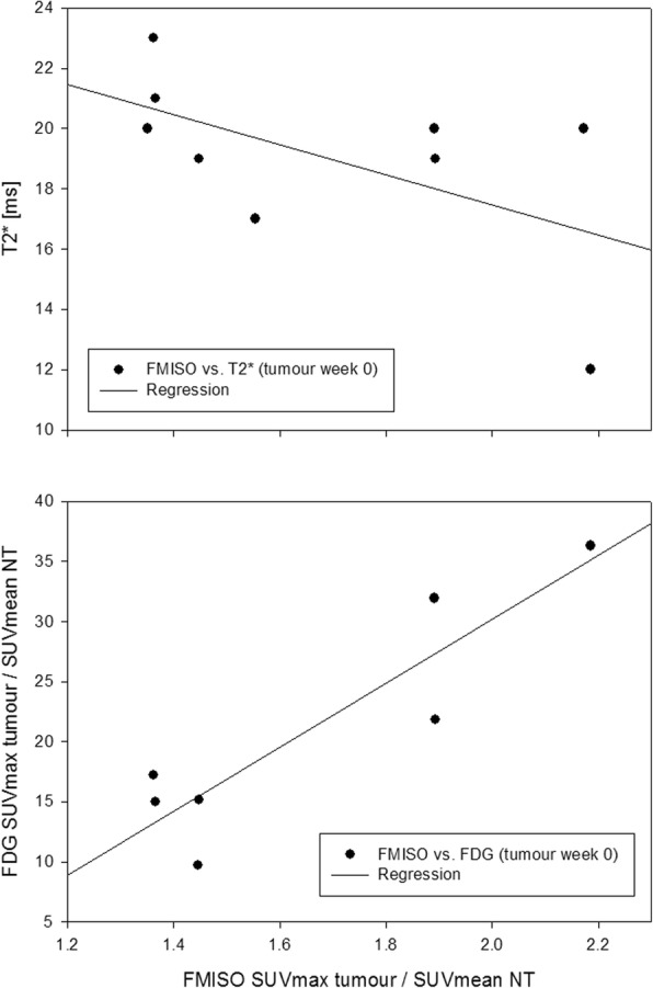
During RCT, marked reduction of tumour hypoxia on FMISO-PET was observed (T, LN), while mean T2* did not change significantly. At baseline, mean T2* values within HSV-T (15 ± 5 ms) were smaller compared to nonHSV-T (18 ± 3 ms; p = 0.051), whereas FDG SUVmean (12 ± 6) was significantly higher for HSV-T (12 ± 6) than for nonHSV-T (6 ± 3; p = 0.026) and higher for HSV-LN (10 ± 4) than for nonHSV-LN (5 ± 2; p ≤ 0.011). Correlation between FMISO PET and FDG PET was higher than between FMSIO PET and T2* (R for GTV-T (FMISO/FDG) = 0.81, R for GTV-T (FMISO/T2*) = 0.32).
Marked reduction of tumour hypoxia between week 0, 2 and 5 found on FMISO PET was not accompanied by a significant T2change within GTVs over time. These results suggest a relation between tumour oxygenation status and T2 at baseline, but no simple correlation over time. Therefore, caution is warranted when using T2* as a substitute for FMISO-PET to monitor tumour hypoxia during RCT in HNSCC patients.
DRKS, DRKS00003830 . Registered 23.04.2012.
评估放化疗(RCT)对拟议肿瘤缺氧标志物横向弛豫时间(T2*)的影响,并分析 T2*与 F-间位碘代苯甲脒正电子发射断层扫描/计算机断层扫描(FMISO-PET)和 F-氟脱氧葡萄糖正电子发射断层扫描/计算机断层扫描(FDG-PET)之间的关系。
10 名接受头颈部鳞状细胞癌(HNSCC)根治性 RCT 的患者在治疗期间的第 0、2 和 5 周接受重复 FMISO 和 3 Tesla T2*-加权 MRI,以及基线 FDG-PET。生成肿瘤(T)、淋巴结(LN)和缺氧亚体积(HSV,基于 FMISO-PET)以及互补非缺氧亚体积(nonHSV)的大体肿瘤体积(GTV)。确定 T2*和 FDG SUVmean 的平均值。
在 RCT 期间,观察到 FMISO-PET 上肿瘤缺氧的显著减少(T,LN),而 T2的平均值没有显著变化。在基线时,HSV-T 内的平均 T2值(15±5 ms)小于非 HSV-T(18±3 ms;p=0.051),而 HSV-T 的 FDG SUVmean(12±6)显著高于非 HSV-T(6±3;p=0.026)和 HSV-LN(10±4)高于非 HSV-LN(5±2;p≤0.011)。FMISO PET 和 FDG PET 之间的相关性高于 FMISO PET 和 T2之间的相关性(GTV-T(FMISO/FDG)的 R 值为 0.81,GTV-T(FMISO/T2)的 R 值为 0.32)。
在 FMISO PET 上发现的 0、2 和 5 周之间肿瘤缺氧的显著减少并未伴随着 GTV 内 T2随时间的显著变化。这些结果表明,肿瘤氧合状态与基线时的 T2之间存在关系,但随时间没有简单的相关性。因此,在使用 T2*替代 FMISO-PET 来监测 HNSCC 患者 RCT 期间的肿瘤缺氧时,需要谨慎。
DRKS,DRKS00003830。注册于 2012 年 4 月 23 日。