Department of Biomedical and Biotechnological Sciences, University of Catania, via S. Sofia 97, 95123 Catania, Italy.
Biomolecules. 2021 Mar 11;11(3):418. doi: 10.3390/biom11030418.
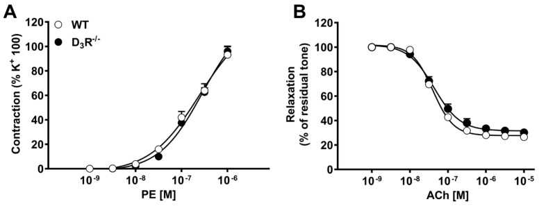
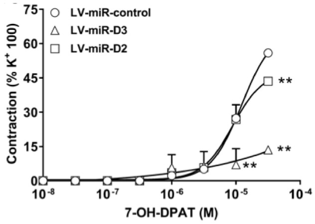
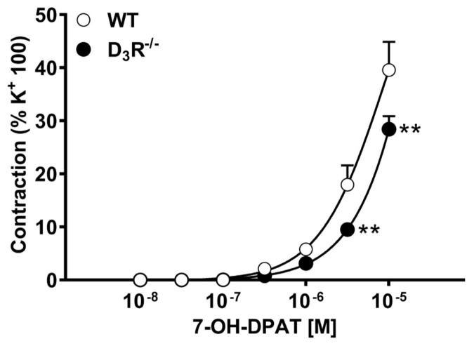
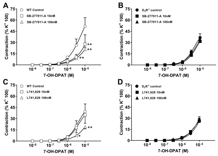
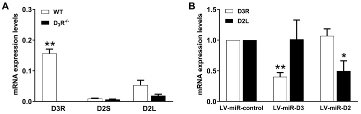
Dopamine receptors (DRs) are generally considered as mediators of vasomotor functions. However, when used in pharmacological studies, dopamine and/or DR agonists may not discriminate among different DR subtypes and may even stimulate alpha1 and beta-adrenoceptors. Here, we tested the hypothesis that D2R and/or D3R may specifically induce vasoconstriction in isolated mouse aorta. Aorta, isolated from wild-type (WT) and D3R/ mice, was mounted in a wire myograph and challenged with cumulative concentrations of phenylephrine (PE), acetylcholine (ACh), and the D3R agonist 7-hydrxy-N,N-dipropyl-2-aminotetralin (7-OH-DPAT), with or without the D2R antagonist L741,626 and the D3R antagonist SB-277011-A. The vasoconstriction to PE and the vasodilatation to ACh were not different in WT and D3R/; in contrast, the contractile responses to 7-OH-DPAT were significantly weaker in D3R/, though not abolished. L741,626 did not change the contractile response induced by 7-OH-DPAT in WT or in D3R/, whereas SB-277011-A significantly reduced it in WT but did not in D3R/. D3R mRNA (assessed by qPCR) was about 5-fold more abundant than D2R mRNA in aorta from WT and undetectable in aorta from D3R/. Following transduction with lentivirus (72-h incubation) delivering synthetic microRNAs to specifically inactivate D2R (LV-miR-D2) or D3R (LV-miR-D3), the contractile response to 7-OH-DPAT was unaffected by LV-miR-D2, while it was significantly reduced by LV-miR-D3. These data indicate that, at least in mouse aorta, D3R stimulation induces vasoconstriction, while D2R stimulation does not. This is consistent with the higher expression level of D3R. The residual vasoconstriction elicited by high concentration D3R agonist in D3R/ and/or in the presence of D3R antagonist is likely to be unrelated to DRs.
多巴胺受体(DRs)通常被认为是血管运动功能的介质。然而,当在药理学研究中使用时,多巴胺和/或 DR 激动剂可能无法区分不同的 DR 亚型,甚至可能刺激 alpha1 和 beta-肾上腺素受体。在这里,我们测试了以下假设:D2R 和/或 D3R 可能特异性地诱导分离的小鼠主动脉收缩。从野生型(WT)和 D3R/小鼠中分离出的主动脉,被安装在一个金属丝肌动描记器中,并使用累积浓度的苯肾上腺素(PE)、乙酰胆碱(ACh)和 D3R 激动剂 7-羟基-N,N-二丙基-2-氨基四氢萘(7-OH-DPAT)进行挑战,同时使用或不使用 D2R 拮抗剂 L741,626 和 D3R 拮抗剂 SB-277011-A。PE 引起的血管收缩和 ACh 引起的血管舒张在 WT 和 D3R/之间没有差异;相反,7-OH-DPAT 引起的收缩反应在 D3R/中明显减弱,尽管没有被消除。L741,626 没有改变 WT 或 D3R/中 7-OH-DPAT 诱导的收缩反应,而 SB-277011-A 在 WT 中显著降低了它,但在 D3R/中没有。通过 qPCR 评估,D3R mRNA 在 WT 主动脉中的丰度约为 D2R mRNA 的 5 倍,在 D3R/主动脉中无法检测到。在用特异性失活 D2R(LV-miR-D2)或 D3R(LV-miR-D3)的合成 microRNA 转导病毒(72 小时孵育)转导后,7-OH-DPAT 的收缩反应不受 LV-miR-D2 的影响,而受 LV-miR-D3 的显著降低。这些数据表明,至少在小鼠主动脉中,D3R 刺激诱导血管收缩,而 D2R 刺激不诱导。这与 D3R 较高的表达水平一致。在 D3R/和/或存在 D3R 拮抗剂的情况下,高浓度 D3R 激动剂引起的残留血管收缩可能与 DR 无关。