Université Paris Est Creteil, INSERM, IMRB, F-94010 Creteil, France.
EFS, IMRB, F-94010 Creteil, France.
Cells. 2021 Mar 28;10(4):744. doi: 10.3390/cells10040744.
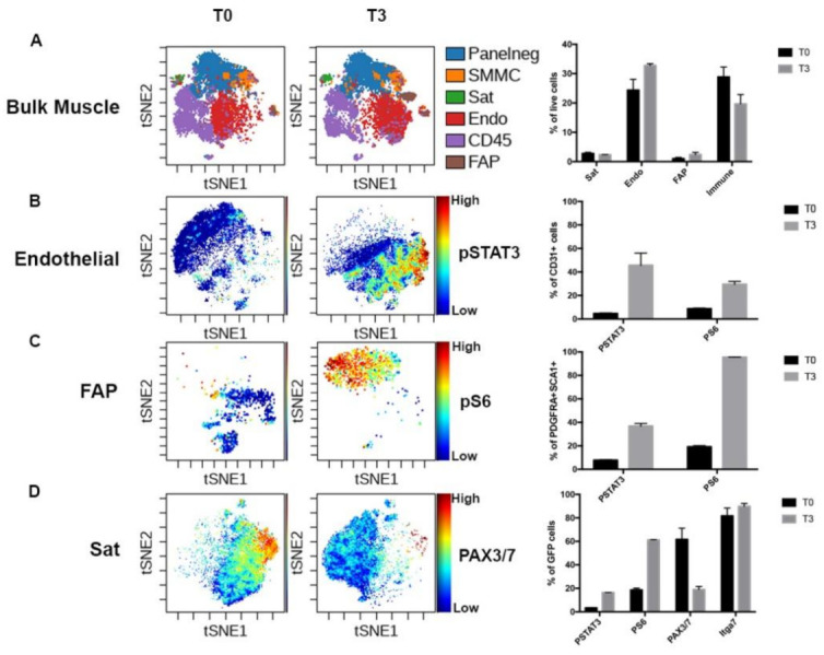
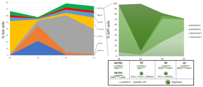
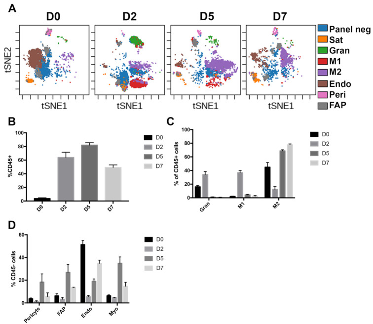
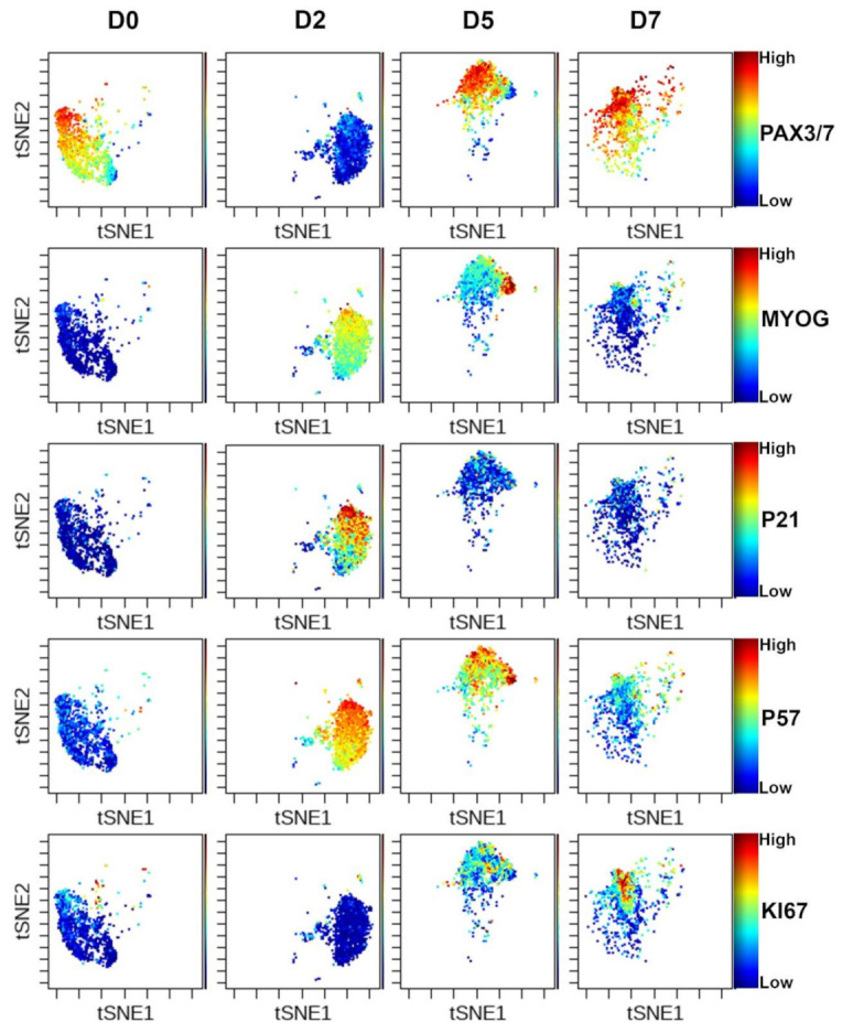
Skeletal muscle is one of the only mammalian tissues capable of rapid and efficient regeneration after trauma or in pathological conditions. Skeletal muscle regeneration is driven by the muscle satellite cells, the stem cell population in interaction with their niche. Upon injury, muscle fibers undergo necrosis and muscle stem cells activate, proliferate and fuse to form new myofibers. In addition to myogenic cell populations, interaction with other cell types such as inflammatory cells, mesenchymal (fibroadipogenic progenitors-FAPs, pericytes) and vascular (endothelial) lineages are important for efficient muscle repair. While the role of the distinct populations involved in skeletal muscle regeneration is well characterized, the quantitative changes in the muscle stem cell and niche during the regeneration process remain poorly characterized. We have used mass cytometry to follow the main muscle cell types (muscle stem cells, vascular, mesenchymal and immune cell lineages) during early activation and over the course of muscle regeneration at D0, D2, D5 and D7 compared with uninjured muscles. Early activation induces a number of rapid changes in the proteome of multiple cell types. Following the induction of damage, we observe a drastic loss of myogenic, vascular and mesenchymal cell lineages while immune cells invade the damaged tissue to clear debris and promote muscle repair. Immune cells constitute up to 80% of the mononuclear cells 5 days post-injury. We show that muscle stem cells are quickly activated in order to form new myofibers and reconstitute the quiescent muscle stem cell pool. In addition, our study provides a quantitative analysis of the various myogenic populations during muscle repair. : We have developed a mass cytometry panel to investigate the dynamic nature of muscle regeneration at a single-cell level. Using our panel, we have identified early changes in the proteome of stressed satellite and niche cells. We have also quantified changes in the major cell types of skeletal muscle during regeneration and analyzed myogenic transcription factor expression in satellite cells throughout this process. Our results highlight the progressive dynamic shifts in cell populations and the distinct states of muscle stem cells adopted during skeletal muscle regeneration. Our findings give a deeper understanding of the cellular and molecular aspects of muscle regeneration.
骨骼肌是哺乳动物组织中唯一能够在创伤后或病理条件下快速而有效地再生的组织。骨骼肌再生由肌肉卫星细胞驱动,这些细胞是与它们的龛位相互作用的干细胞群体。受伤后,肌纤维发生坏死,肌肉干细胞激活、增殖并融合形成新的肌纤维。除了成肌细胞群体外,与其他细胞类型(如炎症细胞、间充质(纤维脂肪前体-FAPs、周细胞)和血管(内皮)谱系)的相互作用对于有效的肌肉修复也很重要。虽然参与骨骼肌再生的不同群体的作用已经得到很好的描述,但在再生过程中肌肉干细胞和龛位的定量变化仍知之甚少。我们使用质谱流式细胞术在 D0、D2、D5 和 D7 与未受伤肌肉相比,在早期激活和整个肌肉再生过程中跟踪主要的肌肉细胞类型(肌肉干细胞、血管、间充质和免疫细胞谱系)。早期激活诱导多种细胞类型的蛋白质组发生快速变化。在诱导损伤后,我们观察到成肌细胞、血管和间充质细胞谱系的急剧丧失,而免疫细胞浸润受损组织以清除碎片并促进肌肉修复。免疫细胞在损伤后 5 天构成单核细胞的 80%。我们表明,肌肉干细胞迅速激活以形成新的肌纤维并重建静止的肌肉干细胞池。此外,我们的研究提供了肌肉修复过程中各种成肌细胞群体的定量分析。我们开发了一种质谱流式细胞术面板来在单细胞水平上研究肌肉再生的动态特性。使用我们的面板,我们确定了应激卫星细胞和龛位细胞蛋白质组的早期变化。我们还量化了再生过程中骨骼肌主要细胞类型的变化,并分析了整个过程中成肌转录因子在卫星细胞中的表达。我们的结果突出了细胞群体的渐进动态变化以及骨骼肌再生过程中肌肉干细胞采用的不同状态。我们的发现更深入地了解了肌肉再生的细胞和分子方面。