• 文献检索
  • 文档翻译
  • 深度研究
  • 学术资讯
  • Suppr Zotero 插件Zotero 插件
  • 邀请有礼
  • 套餐&价格
  • 历史记录
应用&插件
Suppr Zotero 插件Zotero 插件浏览器插件Mac 客户端Windows 客户端微信小程序
定价
高级版会员购买积分包购买API积分包
服务
文献检索文档翻译深度研究API 文档MCP 服务
关于我们
关于 Suppr公司介绍联系我们用户协议隐私条款
关注我们

Suppr 超能文献

核心技术专利:CN118964589B侵权必究
粤ICP备2023148730 号-1Suppr @ 2026

文献检索

告别复杂PubMed语法,用中文像聊天一样搜索,搜遍4000万医学文献。AI智能推荐,让科研检索更轻松。

立即免费搜索

文件翻译

保留排版,准确专业,支持PDF/Word/PPT等文件格式,支持 12+语言互译。

免费翻译文档

深度研究

AI帮你快速写综述,25分钟生成高质量综述,智能提取关键信息,辅助科研写作。

立即免费体验

用于微通道血流研究的手动和自动图像分析分割方法

Manual and Automatic Image Analysis Segmentation Methods for Blood Flow Studies in Microchannels.

作者信息

Carvalho Violeta, Gonçalves Inês M, Souza Andrews, Souza Maria S, Bento David, Ribeiro João E, Lima Rui, Pinho Diana

机构信息

Mechanical Engineering and Resource Sustainability Center (MEtRICs), Mechanical Engineering Department, University of Minho, 4800-058 Guimarães, Portugal.

Instituto Superior Técnico, Universidade de Lisboa, Av. Rovisco Pais, 1049-001 Lisboa, Portugal.

出版信息

Micromachines (Basel). 2021 Mar 18;12(3):317. doi: 10.3390/mi12030317.

DOI:10.3390/mi12030317
PMID:33803615
原文链接:https://pmc.ncbi.nlm.nih.gov/articles/PMC8002955/
Abstract

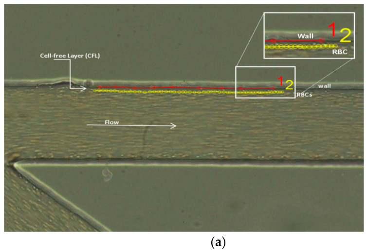
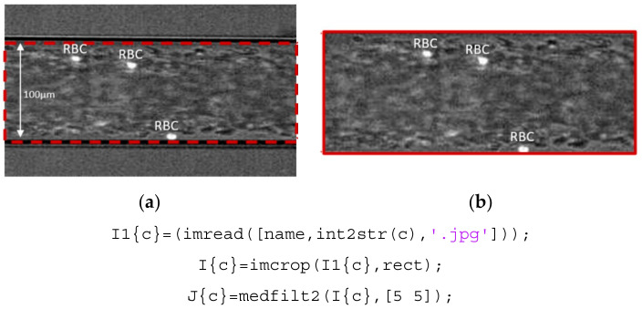
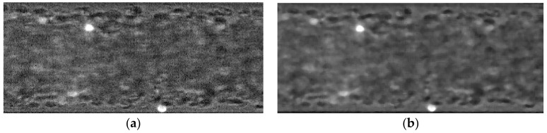
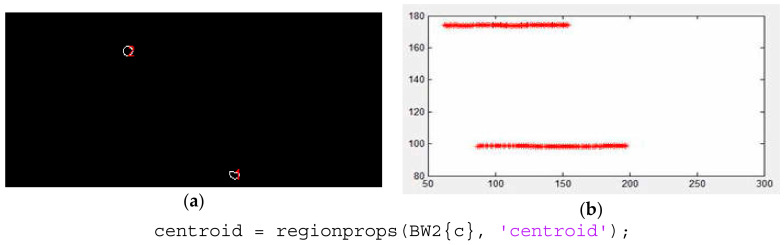
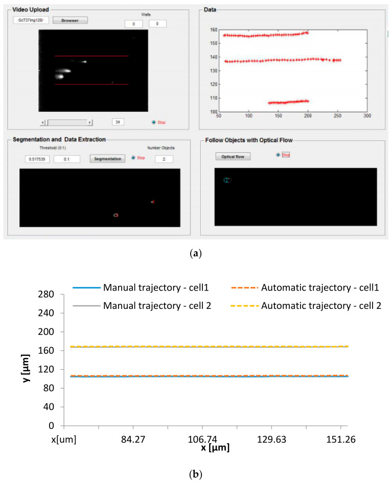
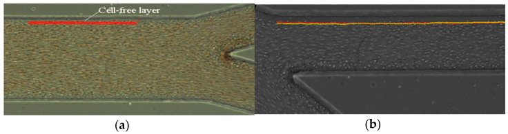
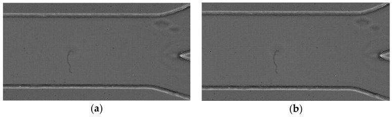
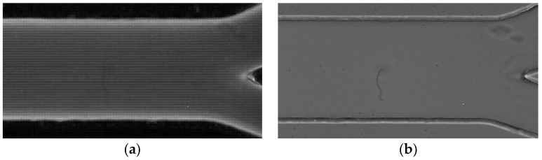
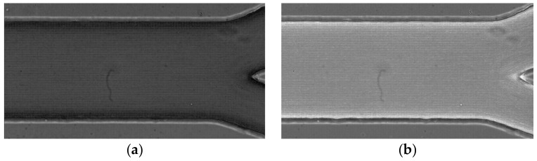
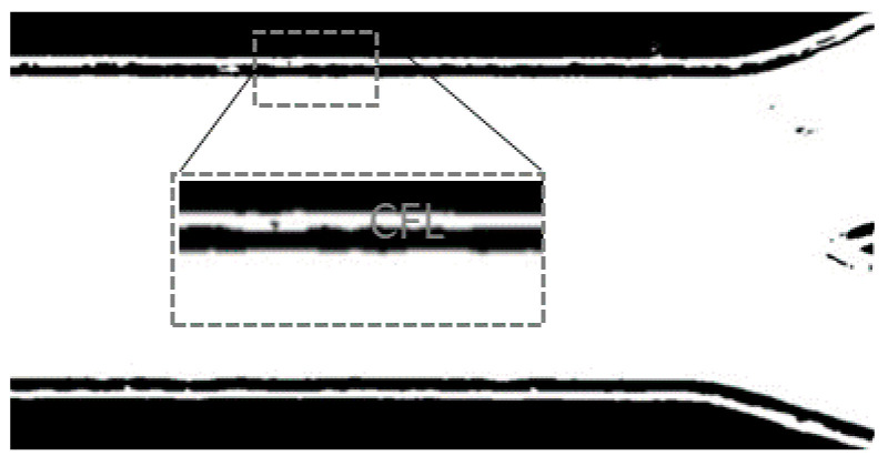
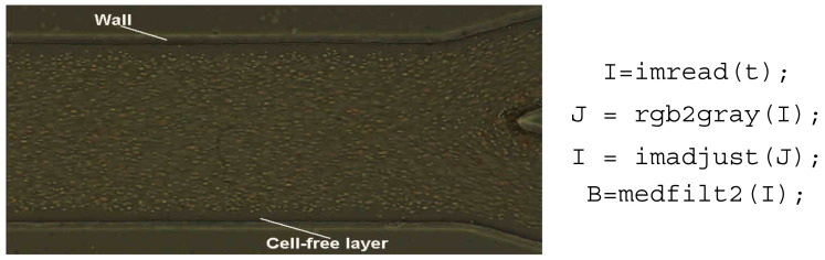
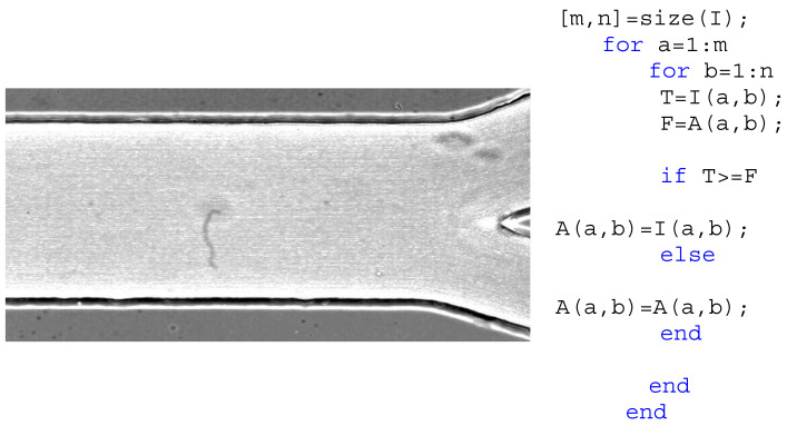
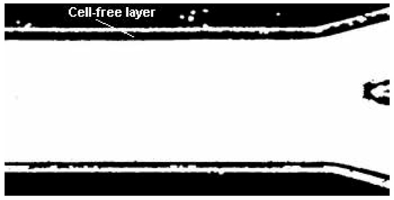
In blood flow studies, image analysis plays an extremely important role to examine raw data obtained by high-speed video microscopy systems. This work shows different ways to process the images which contain various blood phenomena happening in microfluidic devices and in microcirculation. For this purpose, the current methods used for tracking red blood cells (RBCs) flowing through a glass capillary and techniques to measure the cell-free layer thickness in different kinds of microchannels will be presented. Most of the past blood flow experimental data have been collected and analyzed by means of manual methods, that can be extremely reliable, but they are highly time-consuming, user-intensive, repetitive, and the results can be subjective to user-induced errors. For this reason, it is crucial to develop image analysis methods able to obtain the data automatically. Concerning automatic image analysis methods for individual RBCs tracking and to measure the well known microfluidic phenomena cell-free layer, two developed methods are presented and discussed in order to demonstrate their feasibility to obtain accurate data acquisition in such studies. Additionally, a comparison analysis between manual and automatic methods was performed.

摘要

在血流研究中,图像分析对于检查高速视频显微镜系统获取的原始数据起着极其重要的作用。这项工作展示了处理包含微流体装置和微循环中发生的各种血液现象的图像的不同方法。为此,将介绍目前用于跟踪流经玻璃毛细管的红细胞(RBC)的方法以及测量不同类型微通道中无细胞层厚度的技术。过去的大多数血流实验数据都是通过手动方法收集和分析的,这些方法可能非常可靠,但耗时极长、人力密集、重复性高,而且结果可能会受到用户导致的误差的影响。因此,开发能够自动获取数据的图像分析方法至关重要。关于用于单个红细胞跟踪以及测量著名的微流体现象无细胞层的自动图像分析方法,介绍并讨论了两种已开发的方法,以证明它们在此类研究中获取准确数据采集的可行性。此外,还对手动方法和自动方法进行了比较分析。

https://cdn.ncbi.nlm.nih.gov/pmc/blobs/cd0b/8002955/31dc75426f08/micromachines-12-00317-g017.jpg
https://cdn.ncbi.nlm.nih.gov/pmc/blobs/cd0b/8002955/93d5f9344168/micromachines-12-00317-g001a.jpg
https://cdn.ncbi.nlm.nih.gov/pmc/blobs/cd0b/8002955/94bc04419ef4/micromachines-12-00317-g002.jpg
https://cdn.ncbi.nlm.nih.gov/pmc/blobs/cd0b/8002955/ab47ed2d5132/micromachines-12-00317-g003.jpg
https://cdn.ncbi.nlm.nih.gov/pmc/blobs/cd0b/8002955/8a77c2a4fce6/micromachines-12-00317-g004.jpg
https://cdn.ncbi.nlm.nih.gov/pmc/blobs/cd0b/8002955/1e630fe4dda8/micromachines-12-00317-g005.jpg
https://cdn.ncbi.nlm.nih.gov/pmc/blobs/cd0b/8002955/1ff60a27fa9c/micromachines-12-00317-g006.jpg
https://cdn.ncbi.nlm.nih.gov/pmc/blobs/cd0b/8002955/3588d20d530d/micromachines-12-00317-g007.jpg
https://cdn.ncbi.nlm.nih.gov/pmc/blobs/cd0b/8002955/79ef636aa2bd/micromachines-12-00317-g008.jpg
https://cdn.ncbi.nlm.nih.gov/pmc/blobs/cd0b/8002955/10b7db1db6e3/micromachines-12-00317-g009.jpg
https://cdn.ncbi.nlm.nih.gov/pmc/blobs/cd0b/8002955/e9574f9da088/micromachines-12-00317-g010.jpg
https://cdn.ncbi.nlm.nih.gov/pmc/blobs/cd0b/8002955/66d62f1dd7fa/micromachines-12-00317-g011.jpg
https://cdn.ncbi.nlm.nih.gov/pmc/blobs/cd0b/8002955/bd90d26db7f6/micromachines-12-00317-g012.jpg
https://cdn.ncbi.nlm.nih.gov/pmc/blobs/cd0b/8002955/b4914e1f0d0f/micromachines-12-00317-g013.jpg
https://cdn.ncbi.nlm.nih.gov/pmc/blobs/cd0b/8002955/13516d268b55/micromachines-12-00317-g014.jpg
https://cdn.ncbi.nlm.nih.gov/pmc/blobs/cd0b/8002955/9565e30f819a/micromachines-12-00317-g015.jpg
https://cdn.ncbi.nlm.nih.gov/pmc/blobs/cd0b/8002955/412b2907c030/micromachines-12-00317-g016.jpg
https://cdn.ncbi.nlm.nih.gov/pmc/blobs/cd0b/8002955/31dc75426f08/micromachines-12-00317-g017.jpg

相似文献

1
Manual and Automatic Image Analysis Segmentation Methods for Blood Flow Studies in Microchannels.用于微通道血流研究的手动和自动图像分析分割方法
Micromachines (Basel). 2021 Mar 18;12(3):317. doi: 10.3390/mi12030317.
2
Automatic tracking of labeled red blood cells in microchannels.微通道中标记红细胞的自动跟踪。
Int J Numer Method Biomed Eng. 2013 Sep;29(9):977-87. doi: 10.1002/cnm.2526. Epub 2012 Nov 12.
3
Red blood cells radial dispersion in blood flowing through microchannels: The role of temperature.红细胞在流经微通道的血液中的径向扩散:温度的作用。
J Biomech. 2016 Jul 26;49(11):2293-2298. doi: 10.1016/j.jbiomech.2015.11.037. Epub 2015 Nov 28.
4
Semi-automated red blood cell core detection in blood micro-flow.血液微流中红细胞的半自动核心检测
Microvasc Res. 2023 May;147:104496. doi: 10.1016/j.mvr.2023.104496. Epub 2023 Feb 3.
5
Assessment of the Deformability and Velocity of Healthy and Artificially Impaired Red Blood Cells in Narrow Polydimethylsiloxane (PDMS) Microchannels.在狭窄的聚二甲基硅氧烷(PDMS)微通道中对健康及人工损伤红细胞的变形能力和速度进行评估。
Micromachines (Basel). 2018 Aug 2;9(8):384. doi: 10.3390/mi9080384.
6
Particle tracking for the assessment of microcirculatory perfusion.用于评估微循环灌注的粒子追踪技术。
Physiol Meas. 2017 Feb;38(2):358-373. doi: 10.1088/1361-6579/aa56ab. Epub 2017 Jan 4.
7
Deformation of Red Blood Cells, Air Bubbles, and Droplets in Microfluidic Devices: Flow Visualizations and Measurements.微流控装置中红细胞、气泡和液滴的变形:流动可视化与测量
Micromachines (Basel). 2018 Mar 27;9(4):151. doi: 10.3390/mi9040151.
8
Automatic MRI-based Three-dimensional Models of Hip Cartilage Provide Improved Morphologic and Biochemical Analysis.基于 MRI 的髋关节软骨自动三维模型可提高形态学和生物化学分析的效果。
Clin Orthop Relat Res. 2019 May;477(5):1036-1052. doi: 10.1097/CORR.0000000000000755.
9
Flow visualization tools for image analysis of capillary networks.用于毛细血管网络图像分析的流动可视化工具。
Microcirculation. 2004 Jan-Feb;11(1):39-54. doi: 10.1080/10739680490266171.
10
Microfluidic interactions between red blood cells and drug carriers by image analysis techniques.通过图像分析技术研究红细胞与药物载体之间的微流体相互作用。
Med Eng Phys. 2016 Jan;38(1):17-23. doi: 10.1016/j.medengphy.2015.10.005. Epub 2015 Dec 2.

引用本文的文献

1
iCLOTS: open-source, artificial intelligence-enabled software for analyses of blood cells in microfluidic and microscopy-based assays.iCLOTS:开源的人工智能软件,用于分析微流控和基于显微镜的检测中的血细胞。
Nat Commun. 2023 Aug 18;14(1):5022. doi: 10.1038/s41467-023-40522-4.
2
Normalization of Blood Viscosity According to the Hematocrit and the Shear Rate.根据血细胞比容和剪切速率对血液粘度进行标准化。
Micromachines (Basel). 2022 Feb 24;13(3):357. doi: 10.3390/mi13030357.
3
Editorial for the Special Issue on Micro/Nano Devices for Blood Analysis, Volume II.

本文引用的文献

1
Measuring wall shear stress distribution in the carotid artery in an African population: Computational fluid dynamics versus ultrasound doppler velocimetry.测量非洲人群颈动脉壁面剪应力分布:计算流体动力学与超声多普勒测速法的对比
Radiography (Lond). 2021 May;27(2):581-588. doi: 10.1016/j.radi.2020.11.018. Epub 2020 Dec 13.
2
Organ-on-a-Chip: A Preclinical Microfluidic Platform for the Progress of Nanomedicine.器官芯片:推进纳米医学的临床前微流控平台。
Small. 2020 Dec;16(51):e2003517. doi: 10.1002/smll.202003517. Epub 2020 Nov 25.
3
Whole blood viscosity and red blood cell adhesion: Potential biomarkers for targeted and curative therapies in sickle cell disease.
《血液分析微纳器件特刊第二卷》社论
Micromachines (Basel). 2022 Jan 31;13(2):244. doi: 10.3390/mi13020244.
4
Microfluidics Approach to the Mechanical Properties of Red Blood Cell Membrane and Their Effect on Blood Rheology.用于研究红细胞膜力学特性及其对血液流变学影响的微流控方法
Membranes (Basel). 2022 Feb 13;12(2):217. doi: 10.3390/membranes12020217.
5
Properties and Applications of PDMS for Biomedical Engineering: A Review.聚二甲基硅氧烷在生物医学工程中的特性与应用:综述
J Funct Biomater. 2021 Dec 21;13(1):2. doi: 10.3390/jfb13010002.
全血黏度和红细胞黏附:镰状细胞病靶向和治愈疗法的潜在生物标志物。
Am J Hematol. 2020 Nov;95(11):1246-1256. doi: 10.1002/ajh.25933. Epub 2020 Aug 10.
4
Bubbles Moving in Blood Flow in a Microchannel Network: The Effect on the Local Hematocrit.微通道网络中血流中的气泡:对局部血细胞比容的影响。
Micromachines (Basel). 2020 Mar 26;11(4):344. doi: 10.3390/mi11040344.
5
Evaluation and benchmarking of level set-based three forces via geometric active contours for segmentation of white blood cell nuclei shape.基于水平集的三种力通过几何活动轮廓对白细胞细胞核形状进行分割的评估与基准测试。
Comput Biol Med. 2020 Jan;116:103568. doi: 10.1016/j.compbiomed.2019.103568. Epub 2019 Nov 30.
6
Blood Speckle-Tracking Based on High-Frame Rate Ultrasound Imaging in Pediatric Cardiology.基于高速超声成像的儿科心脏病学中的血斑跟踪。
J Am Soc Echocardiogr. 2020 Apr;33(4):493-503.e5. doi: 10.1016/j.echo.2019.11.003. Epub 2020 Jan 24.
7
4-D Echo-Particle Image Velocimetry in a Left Ventricular Phantom.左心室模型中的 4-D 超声粒子图像测速
Ultrasound Med Biol. 2020 Mar;46(3):805-817. doi: 10.1016/j.ultrasmedbio.2019.11.020. Epub 2020 Jan 8.
8
Deep Learning-Based Super-resolution Ultrasound Speckle Tracking Velocimetry.基于深度学习的超声散斑跟踪速度测量超分辨率技术。
Ultrasound Med Biol. 2020 Mar;46(3):598-609. doi: 10.1016/j.ultrasmedbio.2019.12.002. Epub 2020 Jan 6.
9
Detection of red and white blood cells from microscopic blood images using a region proposal approach.基于区域提议的方法检测显微镜下血图像中的红细胞和白细胞。
Comput Biol Med. 2020 Jan;116:103530. doi: 10.1016/j.compbiomed.2019.103530. Epub 2019 Nov 7.
10
Blood Cells Separation and Sorting Techniques of Passive Microfluidic Devices: From Fabrication to Applications.被动微流控装置的血细胞分离与分选技术:从制造到应用
Micromachines (Basel). 2019 Sep 10;10(9):593. doi: 10.3390/mi10090593.