Durstberger Gerlinde, Nguyen Phuong Quynh, Hohensinner Verena, Pietschmann Peter, Rausch-Fan Xiaohui, Andrukhov Oleh
Division of Conservative Dentistry and Periodontology, University Clinic of Dentistry, Medical University of Vienna, 1090 Vienna, Austria.
Competence Center for Periodontal Research, University Clinic of Dentistry, Medical University of Vienna, 1090 Vienna, Austria.
Medicina (Kaunas). 2021 Mar 15;57(3):269. doi: 10.3390/medicina57030269.
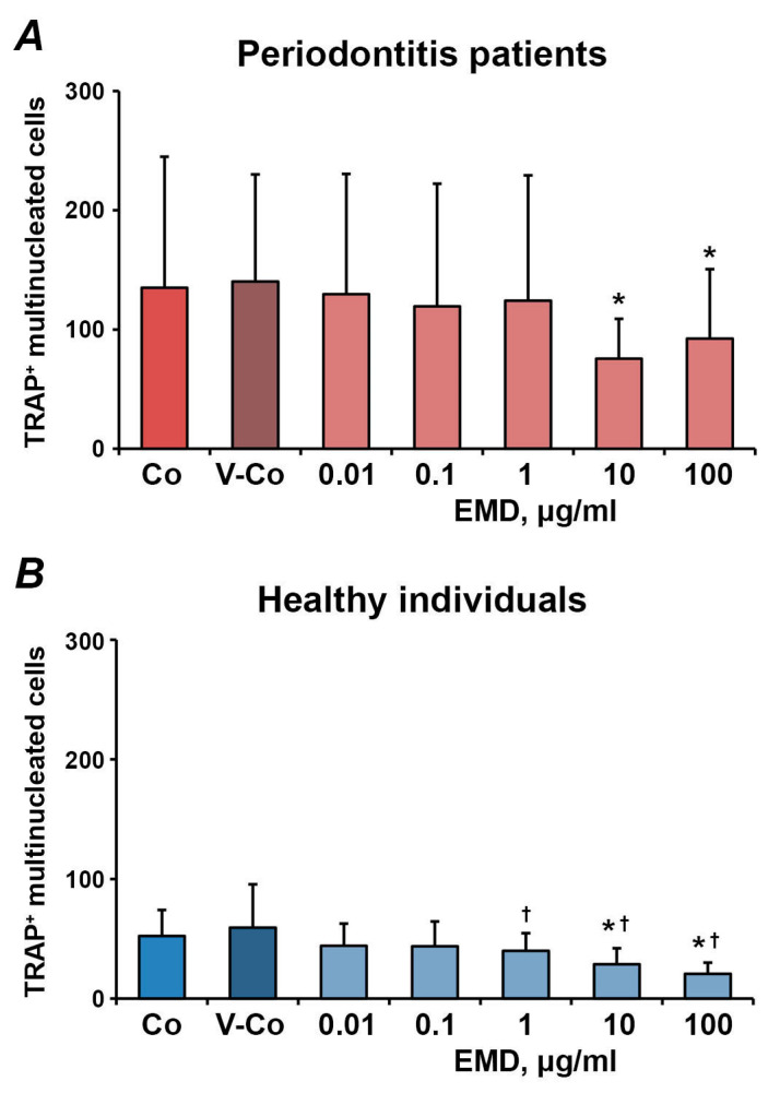
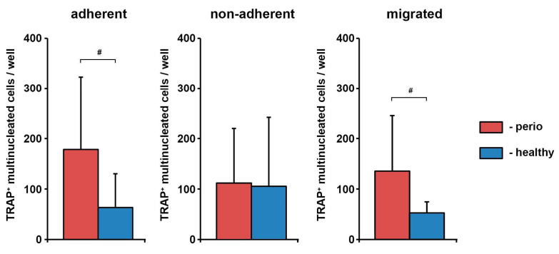
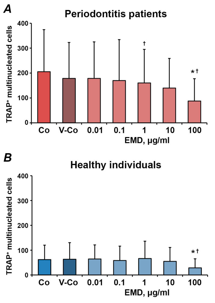
Enamel matrix derivative (EMD) is produced from developing porcine tooth buds and represents a complex of low-molecular-weight hydrophobic enamel proteins. EMD is widely applied in periodontal regeneration. Osteoclasts are multinuclear cells, which are responsible for bone resorption. The precursors of osteoclasts, hematopoietic cells, undergo in vivo the process of transendothelial migration before differentiation. EMD is known to affect the process of osteoclastogenesis, but its effect on human osteoclasts precursors after the interaction with activated endothelium was never studied. Human umbilical vein endothelial cells (HUVECs)s were seeded in transwell inserts with a pore size of 8 µm and pre-activated by TNF-α and IL-1β for 18 h. Peripheral blood mononuclear cells (PBMCs), freshly isolated from 16 periodontitis patients and 16 healthy individuals, were added to pre-activated HUVECs. Adherent, non-adherent and transmigrated cells were collected and differentiated to osteoclasts by the standard protocol in the presence or absence of EMD. The number of osteoclasts was determined by tartrate-resistant acid phosphatase staining. PBMCs isolated from periodontitis patients have formed a significantly higher osteoclast number compared to PBMCs isolated from healthy individuals ( < 0.05). EMD induced concentration-dependent inhibition of osteoclast formation from PBMCs. This was true for the different PBMC fractions isolated from both healthy individuals and periodontitis patients. Our data show that EMD inhibits the formation and activity of osteoclasts differentiated from the progenitor cells after the interaction with activated endothelium. This might be associated with bone resorption inhibition and supporting bone regeneration in the frame of periodontal therapy.
釉基质衍生物(EMD)由发育中的猪牙胚产生,是一种低分子量疏水釉质蛋白复合物。EMD广泛应用于牙周再生。破骨细胞是多核细胞,负责骨吸收。破骨细胞的前体细胞,即造血细胞,在分化前在体内经历跨内皮迁移过程。已知EMD会影响破骨细胞生成过程,但从未研究过其与活化内皮细胞相互作用后对人破骨细胞前体细胞的影响。将人脐静脉内皮细胞(HUVECs)接种在孔径为8 µm的Transwell小室中,并用TNF-α和IL-1β预激活18小时。将从16名牙周炎患者和16名健康个体中新鲜分离的外周血单核细胞(PBMCs)添加到预激活的HUVECs中。收集贴壁、非贴壁和迁移的细胞,并在有或没有EMD的情况下通过标准方案分化为破骨细胞。通过抗酒石酸酸性磷酸酶染色确定破骨细胞的数量。与从健康个体中分离的PBMCs相比,从牙周炎患者中分离的PBMCs形成的破骨细胞数量明显更高(<0.05)。EMD诱导PBMCs破骨细胞形成的浓度依赖性抑制。从健康个体和牙周炎患者中分离的不同PBMC组分均是如此。我们的数据表明,EMD抑制与活化内皮细胞相互作用后祖细胞分化形成的破骨细胞的形成和活性。这可能与牙周治疗框架内的骨吸收抑制和支持骨再生有关。