• 文献检索
  • 文档翻译
  • 深度研究
  • 学术资讯
  • Suppr Zotero 插件Zotero 插件
  • 邀请有礼
  • 套餐&价格
  • 历史记录
应用&插件
Suppr Zotero 插件Zotero 插件浏览器插件Mac 客户端Windows 客户端微信小程序
定价
高级版会员购买积分包购买API积分包
服务
文献检索文档翻译深度研究API 文档MCP 服务
关于我们
关于 Suppr公司介绍联系我们用户协议隐私条款
关注我们

Suppr 超能文献

核心技术专利:CN118964589B侵权必究
粤ICP备2023148730 号-1Suppr @ 2026

文献检索

告别复杂PubMed语法,用中文像聊天一样搜索,搜遍4000万医学文献。AI智能推荐,让科研检索更轻松。

立即免费搜索

文件翻译

保留排版,准确专业,支持PDF/Word/PPT等文件格式,支持 12+语言互译。

免费翻译文档

深度研究

AI帮你快速写综述,25分钟生成高质量综述,智能提取关键信息,辅助科研写作。

立即免费体验

通过细胞骨架以及源自或汇聚于细胞骨架的信号传导过程对关节软骨细胞表型的调控:迈向对肌动蛋白动力学与软骨形成功能之间交叉点的全新理解

Articular Chondrocyte Phenotype Regulation through the Cytoskeleton and the Signaling Processes That Originate from or Converge on the Cytoskeleton: Towards a Novel Understanding of the Intersection between Actin Dynamics and Chondrogenic Function.

作者信息

Lauer Jasmin C, Selig Mischa, Hart Melanie L, Kurz Bodo, Rolauffs Bernd

机构信息

G.E.R.N. Research Center for Tissue Replacement, Regeneration & Neogenesis, Department of Orthopedics and Trauma Surgery, Faculty of Medicine, Medical Center-Albert-Ludwigs-University of Freiburg, 79085 Freiburg im Breisgau, Germany.

Faculty of Biology, University of Freiburg, Schaenzlestrasse 1, D-79104 Freiburg, Germany.

出版信息

Int J Mol Sci. 2021 Mar 23;22(6):3279. doi: 10.3390/ijms22063279.

DOI:10.3390/ijms22063279
PMID:33807043
原文链接:https://pmc.ncbi.nlm.nih.gov/articles/PMC8004672/
Abstract

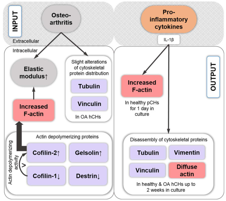
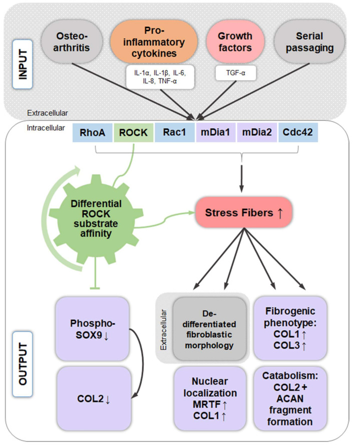
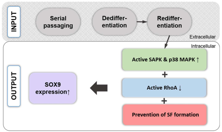
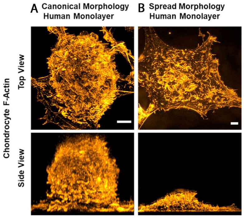
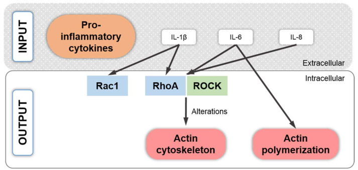
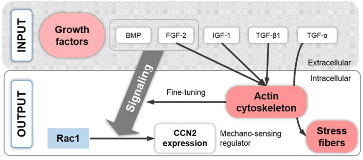
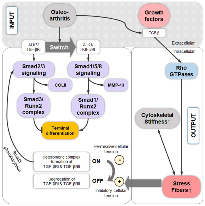
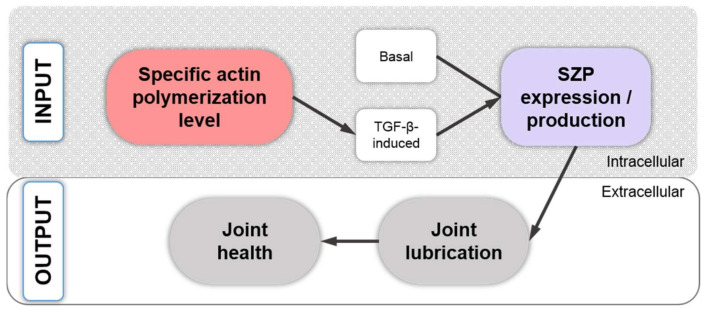
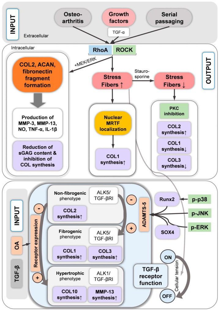
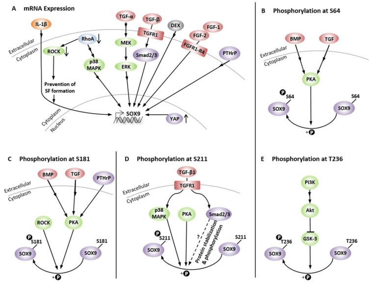
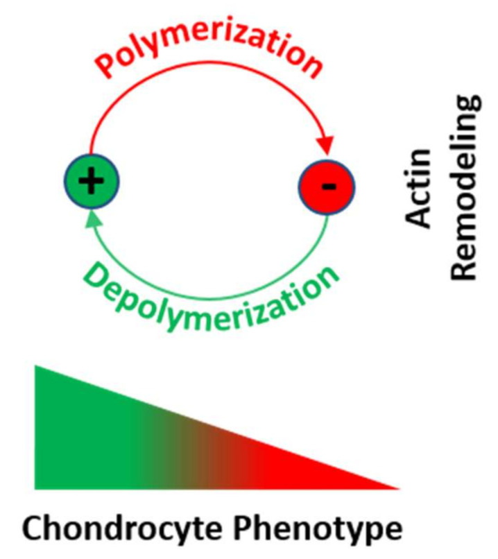
Numerous studies have assembled a complex picture, in which extracellular stimuli and intracellular signaling pathways modulate the chondrocyte phenotype. Because many diseases are mechanobiology-related, this review asked to what extent phenotype regulators control chondrocyte function through the cytoskeleton and cytoskeleton-regulating signaling processes. Such information would generate leverage for advanced articular cartilage repair. Serial passaging, pro-inflammatory cytokine signaling (TNF-α, IL-1α, IL-1β, IL-6, and IL-8), growth factors (TGF-α), and osteoarthritis not only induce dedifferentiation but also converge on RhoA/ROCK/Rac1/mDia1/mDia2/Cdc42 to promote actin polymerization/crosslinking for stress fiber (SF) formation. SF formation takes center stage in phenotype control, as both SF formation and SOX9 phosphorylation for expression are ROCK activity-dependent. Explaining how it is molecularly possible that dedifferentiation induces low expression but high SF formation, this review theorized that, in chondrocyte SOX9, phosphorylation by ROCK might effectively be sidelined in favor of other SF-promoting ROCK substrates, based on a differential ROCK affinity. In turn, actin depolymerization for redifferentiation would "free-up" ROCK to increase expression. Moreover, the actin cytoskeleton regulates expression, modulates COL2/aggrecan fragment generation, and mediates a fibrogenic/catabolic expression profile, highlighting that actin dynamics-regulating processes decisively control the chondrocyte phenotype. This suggests modulating the balance between actin polymerization/depolymerization for therapeutically controlling the chondrocyte phenotype.

摘要

众多研究描绘出了一幅复杂的图景,其中细胞外刺激和细胞内信号通路调节软骨细胞表型。由于许多疾病与机械生物学相关,本综述探讨了表型调节因子在多大程度上通过细胞骨架和细胞骨架调节信号过程来控制软骨细胞功能。此类信息将为高级关节软骨修复提供助力。连续传代、促炎细胞因子信号传导(TNF-α、IL-1α、IL-1β、IL-6和IL-8)、生长因子(TGF-α)以及骨关节炎不仅会诱导去分化,还会汇聚于RhoA/ROCK/Rac1/mDia1/mDia2/Cdc42,以促进肌动蛋白聚合/交联,从而形成应力纤维(SF)。SF形成在表型控制中占据核心地位,因为SF形成和SOX9磷酸化以进行表达均依赖于ROCK活性。为了解释去分化如何在分子层面导致低表达但高SF形成,本综述提出理论,基于不同的ROCK亲和力,在软骨细胞SOX9中,ROCK磷酸化可能有效地被边缘化,转而有利于其他促进SF的ROCK底物。反过来,用于再分化的肌动蛋白解聚将“释放”ROCK以增加表达。此外,肌动蛋白细胞骨架调节表达,调节COL2/聚集蛋白聚糖片段的产生,并介导促纤维化/分解代谢的表达谱,这突出表明调节肌动蛋白动力学的过程决定性地控制着软骨细胞表型。这表明调节肌动蛋白聚合/解聚之间的平衡以治疗性地控制软骨细胞表型。

https://cdn.ncbi.nlm.nih.gov/pmc/blobs/b199/8004672/a6e8fe14f4c3/ijms-22-03279-g014.jpg
https://cdn.ncbi.nlm.nih.gov/pmc/blobs/b199/8004672/1ddef6620704/ijms-22-03279-g005.jpg
https://cdn.ncbi.nlm.nih.gov/pmc/blobs/b199/8004672/243a7152f6b0/ijms-22-03279-g001.jpg
https://cdn.ncbi.nlm.nih.gov/pmc/blobs/b199/8004672/51ea6c028554/ijms-22-03279-g002.jpg
https://cdn.ncbi.nlm.nih.gov/pmc/blobs/b199/8004672/deaf500c10fd/ijms-22-03279-g003.jpg
https://cdn.ncbi.nlm.nih.gov/pmc/blobs/b199/8004672/b29815b0709d/ijms-22-03279-g004.jpg
https://cdn.ncbi.nlm.nih.gov/pmc/blobs/b199/8004672/6252a7fa8286/ijms-22-03279-g006.jpg
https://cdn.ncbi.nlm.nih.gov/pmc/blobs/b199/8004672/a4e357465218/ijms-22-03279-g007.jpg
https://cdn.ncbi.nlm.nih.gov/pmc/blobs/b199/8004672/61642c358962/ijms-22-03279-g008.jpg
https://cdn.ncbi.nlm.nih.gov/pmc/blobs/b199/8004672/01450e6e0962/ijms-22-03279-g009.jpg
https://cdn.ncbi.nlm.nih.gov/pmc/blobs/b199/8004672/fa25ed1a2f15/ijms-22-03279-g010.jpg
https://cdn.ncbi.nlm.nih.gov/pmc/blobs/b199/8004672/30d1f8b18539/ijms-22-03279-g011.jpg
https://cdn.ncbi.nlm.nih.gov/pmc/blobs/b199/8004672/d262b4a2d370/ijms-22-03279-g012.jpg
https://cdn.ncbi.nlm.nih.gov/pmc/blobs/b199/8004672/8c9cbe66de64/ijms-22-03279-g013.jpg
https://cdn.ncbi.nlm.nih.gov/pmc/blobs/b199/8004672/a6e8fe14f4c3/ijms-22-03279-g014.jpg

相似文献

1
Articular Chondrocyte Phenotype Regulation through the Cytoskeleton and the Signaling Processes That Originate from or Converge on the Cytoskeleton: Towards a Novel Understanding of the Intersection between Actin Dynamics and Chondrogenic Function.通过细胞骨架以及源自或汇聚于细胞骨架的信号传导过程对关节软骨细胞表型的调控:迈向对肌动蛋白动力学与软骨形成功能之间交叉点的全新理解
Int J Mol Sci. 2021 Mar 23;22(6):3279. doi: 10.3390/ijms22063279.
2
Interplay between cytoskeletal polymerization and the chondrogenic phenotype in chondrocytes passaged in monolayer culture.单层培养传代软骨细胞中细胞骨架聚合与软骨细胞表型之间的相互作用。
J Anat. 2017 Feb;230(2):234-248. doi: 10.1111/joa.12554. Epub 2016 Nov 3.
3
Adseverin, an actin binding protein, regulates articular chondrocyte phenotype.整合蛋白 Adseverin 调节关节软骨细胞表型。
J Tissue Eng Regen Med. 2019 Aug;13(8):1438-1452. doi: 10.1002/term.2898. Epub 2019 Jun 14.
4
Targeting F-actin stress fibers to suppress the dedifferentiated phenotype in chondrocytes.靶向 F- 肌动蛋白应力纤维抑制软骨细胞去分化表型。
Eur J Cell Biol. 2024 Jun;103(2):151424. doi: 10.1016/j.ejcb.2024.151424. Epub 2024 May 25.
5
Characterization of the chondrocyte actin cytoskeleton in living three-dimensional culture: response to anabolic and catabolic stimuli.活三维培养中软骨细胞肌动蛋白细胞骨架的特征:对合成代谢和分解代谢刺激的反应。
Mol Cell Biomech. 2009 Sep;6(3):135-44.
6
Interference with the contractile machinery of the fibroblastic chondrocyte cytoskeleton induces re-expression of the cartilage phenotype through involvement of PI3K, PKC and MAPKs.干扰成纤维细胞软骨细胞细胞骨架的收缩机制,通过 PI3K、PKC 和 MAPKs 的参与,诱导软骨表型的重新表达。
Exp Cell Res. 2014 Jan 15;320(2):175-87. doi: 10.1016/j.yexcr.2013.11.004. Epub 2013 Nov 15.
7
RhoA/ROCK signaling regulates Sox9 expression and actin organization during chondrogenesis.RhoA/ROCK信号通路在软骨形成过程中调节Sox9表达和肌动蛋白组织。
J Biol Chem. 2005 Mar 25;280(12):11626-34. doi: 10.1074/jbc.M409158200. Epub 2005 Jan 21.
8
The transcriptional activity of Sox9 in chondrocytes is regulated by RhoA signaling and actin polymerization.软骨细胞中Sox9的转录活性受RhoA信号传导和肌动蛋白聚合作用的调控。
Mol Cell Biol. 2009 Aug;29(15):4262-73. doi: 10.1128/MCB.01779-08. Epub 2009 May 26.
9
Flavonoid Compound Icariin Activates Hypoxia Inducible Factor-1α in Chondrocytes and Promotes Articular Cartilage Repair.黄酮类化合物淫羊藿苷激活软骨细胞中的缺氧诱导因子-1α并促进关节软骨修复。
PLoS One. 2016 Feb 3;11(2):e0148372. doi: 10.1371/journal.pone.0148372. eCollection 2016.
10
RhoA/ROCK signaling regulates chondrogenesis in a context-dependent manner.RhoA/ROCK信号通路以一种依赖于背景的方式调节软骨形成。
J Biol Chem. 2006 May 12;281(19):13134-13140. doi: 10.1074/jbc.M509433200. Epub 2006 Mar 24.

引用本文的文献

1
Insights into chondrocyte populations in cartilaginous tissues at the single-cell level.单细胞水平下软骨组织中软骨细胞群体的见解。
Nat Rev Rheumatol. 2025 Jul 10. doi: 10.1038/s41584-025-01275-0.
2
Sarcoglycans Role in Actin Cytoskeleton Dynamics and Cell Adhesion of Human Articular Chondrocytes: New Insights from siRNA-Mediated Gene Silencing.肌聚糖在人关节软骨细胞肌动蛋白细胞骨架动力学和细胞黏附中的作用:来自小干扰RNA介导基因沉默的新见解
Int J Mol Sci. 2025 Jun 15;26(12):5732. doi: 10.3390/ijms26125732.
3
Identification biomarkers and therapeutic targets of disulfidptosis-related in rheumatoid arthritis via bioinformatics, molecular dynamics simulation, and experimental validation.

本文引用的文献

1
An Evidence-Based Systematic Review of Human Knee Post-Traumatic Osteoarthritis (PTOA): Timeline of Clinical Presentation and Disease Markers, Comparison of Knee Joint PTOA Models and Early Disease Implications.基于证据的人体膝关节创伤后骨关节炎(PTOA)系统评价:临床发病时间和疾病标志物、膝关节 PTOA 模型比较和早期疾病影响。
Int J Mol Sci. 2021 Feb 17;22(4):1996. doi: 10.3390/ijms22041996.
2
Twinfilin uncaps filament barbed ends to promote turnover of lamellipodial actin networks.双胎filin 去帽纤维丝端以促进片状伪足 actin 网络的周转率。
Nat Cell Biol. 2021 Feb;23(2):147-159. doi: 10.1038/s41556-020-00629-y. Epub 2021 Feb 8.
3
通过生物信息学、分子动力学模拟和实验验证确定类风湿关节炎中与二硫键连接的细胞程序性坏死相关的生物标志物和治疗靶点。
Sci Rep. 2025 Mar 13;15(1):8779. doi: 10.1038/s41598-025-93656-4.
4
Piezo1 promotes intervertebral disc degeneration through the Ca/F-actin/Yap signaling axis.Piezo1通过Ca/F-肌动蛋白/Yap信号轴促进椎间盘退变。
Mol Med. 2025 Mar 8;31(1):90. doi: 10.1186/s10020-025-01147-z.
5
The role of RhoA-ROCK signaling in benign prostatic hyperplasia: a review.RhoA-ROCK信号通路在良性前列腺增生中的作用:综述
Hum Cell. 2025 Feb 1;38(2):48. doi: 10.1007/s13577-025-01179-x.
6
From polarity to pathology: Decoding the role of cell orientation in osteoarthritis.从极性到病理学:解读细胞取向在骨关节炎中的作用
J Orthop Translat. 2024 Oct 4;49:62-73. doi: 10.1016/j.jot.2024.09.004. eCollection 2024 Nov.
7
Characteristics of immune cells and causal relationship with chondromalacia: A two-sample, bidirectional mendelian randomization study.免疫细胞特征与软骨软化症的因果关系:两样本双向孟德尔随机化研究。
Mol Pain. 2024 Jan-Dec;20:17448069241289962. doi: 10.1177/17448069241289962.
8
Chemically programmed metabolism drives a superior cell fitness for cartilage regeneration.化学编程代谢可提高软骨再生的细胞适应性。
Sci Adv. 2024 Sep 13;10(37):eadp4408. doi: 10.1126/sciadv.adp4408. Epub 2024 Sep 11.
9
Identification and Construction of a Disulfidptosis-Mediated Diagnostic Model and Associated Immune Microenvironment of Osteoarthritis from the Perspective of PPPM.从PPPM视角识别与构建二硫键介导的骨关节炎诊断模型及相关免疫微环境
J Inflamm Res. 2024 Jun 11;17:3753-3770. doi: 10.2147/JIR.S462179. eCollection 2024.
10
Targeting F-actin stress fibers to suppress the dedifferentiated phenotype in chondrocytes.靶向 F- 肌动蛋白应力纤维抑制软骨细胞去分化表型。
Eur J Cell Biol. 2024 Jun;103(2):151424. doi: 10.1016/j.ejcb.2024.151424. Epub 2024 May 25.
Actin dynamics as critical ion channel regulator: ENaC and Piezo in focus.
肌动蛋白动力学作为关键的离子通道调节剂:ENaC 和 Piezo 成为焦点。
Am J Physiol Cell Physiol. 2021 May 1;320(5):C696-C702. doi: 10.1152/ajpcell.00368.2020. Epub 2021 Jan 20.
4
Integrin α2β1 plays an important role in the interaction between human articular cartilage-derived chondrocytes and atelocollagen gel.整合素 α2β1 在人关节软骨来源的软骨细胞与去端肽胶原凝胶的相互作用中发挥重要作用。
Sci Rep. 2021 Jan 19;11(1):1757. doi: 10.1038/s41598-021-81378-2.
5
Proof-of-concept for the detection of early osteoarthritis pathology by clinically applicable endomicroscopy and quantitative AI-supported optical biopsy.通过临床适用的内窥式显微镜和定量人工智能支持的光学活检术检测早期骨关节炎病理的概念验证。
Osteoarthritis Cartilage. 2021 Feb;29(2):269-279. doi: 10.1016/j.joca.2020.10.003. Epub 2020 Nov 19.
6
Roles of CCN2 as a mechano-sensing regulator of chondrocyte differentiation.CCN2作为软骨细胞分化的机械传感调节因子的作用。
Jpn Dent Sci Rev. 2020 Nov;56(1):119-126. doi: 10.1016/j.jdsr.2020.07.001. Epub 2020 Oct 9.
7
Mechanotransduction and Stiffness-Sensing: Mechanisms and Opportunities to Control Multiple Molecular Aspects of Cell Phenotype as a Design Cornerstone of Cell-Instructive Biomaterials for Articular Cartilage Repair.力学转导和刚性感知:控制细胞表型多个分子方面的机制和机遇,作为关节软骨修复细胞指令性生物材料设计基石。
Int J Mol Sci. 2020 Jul 29;21(15):5399. doi: 10.3390/ijms21155399.
8
Safety and efficacy of ripasudil in Japanese patients with glaucoma or ocular hypertension: 12-month interim analysis of ROCK-J, a post-marketing surveillance study.利匹鲁唑治疗日本青光眼或高眼压症患者的安全性和疗效:ROCK-J 上市后监测研究的 12 个月中期分析。
BMC Ophthalmol. 2020 Jul 9;20(1):275. doi: 10.1186/s12886-020-01490-1.
9
Cell response to substrate rigidity is regulated by active and passive cytoskeletal stress.细胞对基质硬度的反应受细胞骨架的主动和被动应力调节。
Proc Natl Acad Sci U S A. 2020 Jun 9;117(23):12817-12825. doi: 10.1073/pnas.1917555117. Epub 2020 May 22.
10
Ripasudil Hydrochloride Hydrate in the Treatment of Glaucoma: Safety, Efficacy, and Patient Selection.盐酸水合利马前列素治疗青光眼:安全性、有效性及患者选择
Clin Ophthalmol. 2020 May 6;14:1229-1236. doi: 10.2147/OPTH.S216907. eCollection 2020.