Department of Pharmaceutics, State Key Laboratory of Nature Medicines, China Pharmaceutical University, Nanjing, People's Republic of China.
Jiangsu Health Vocational College, Nanjing, People's Republic of China.
Int J Nanomedicine. 2021 Mar 26;16:2443-2459. doi: 10.2147/IJN.S289228. eCollection 2021.
Specific modifications to carriers to achieve targeted delivery of chemotherapeutics into malignant tissues are a critical point for efficient diagnosis and therapy. In this case, bovine serum albumin (BSA) was conjugated with cetuximab-valine-citrulline (vc)-doxorubicin (DOX) to target epidermal growth factor receptor (EGFR) and enable the release of drug in EGFR-overexpressed tumor cells.
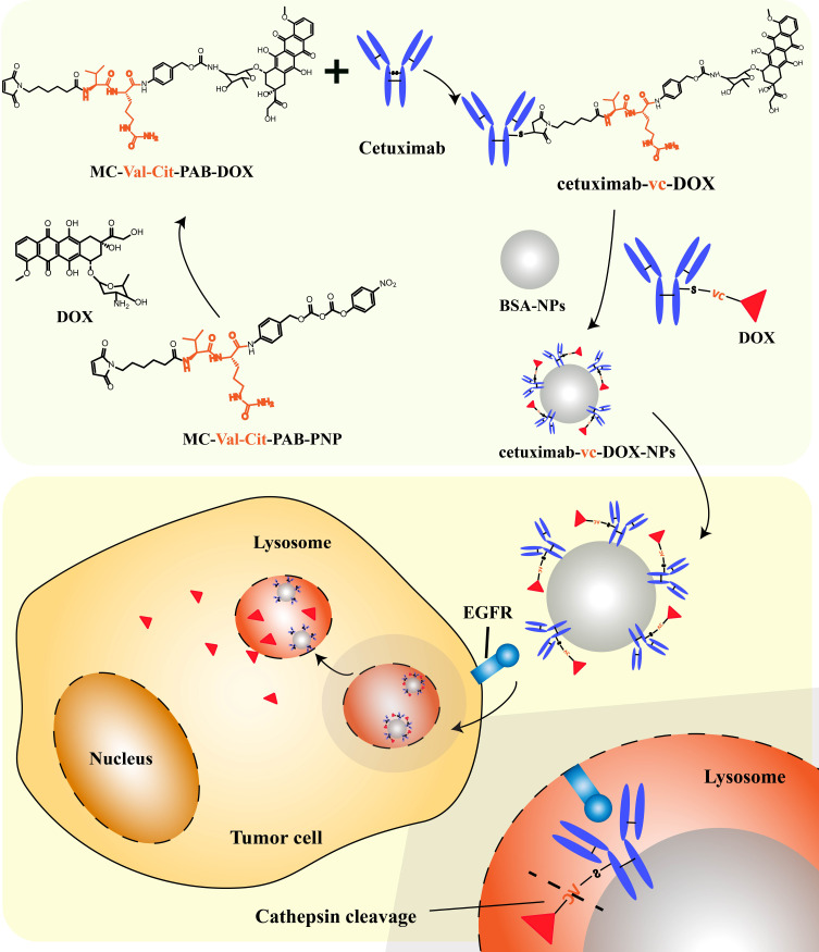
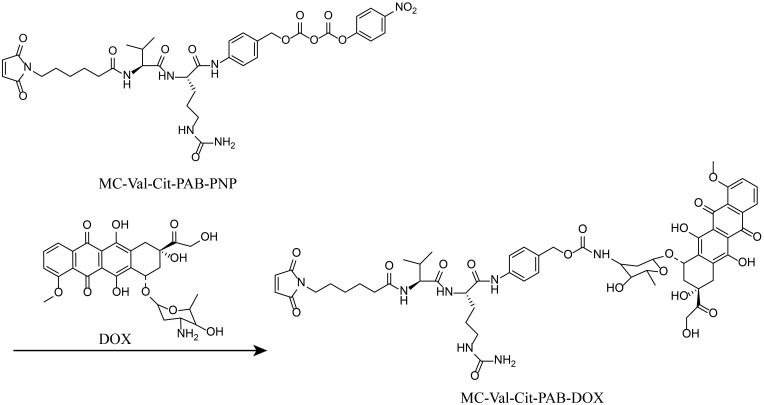
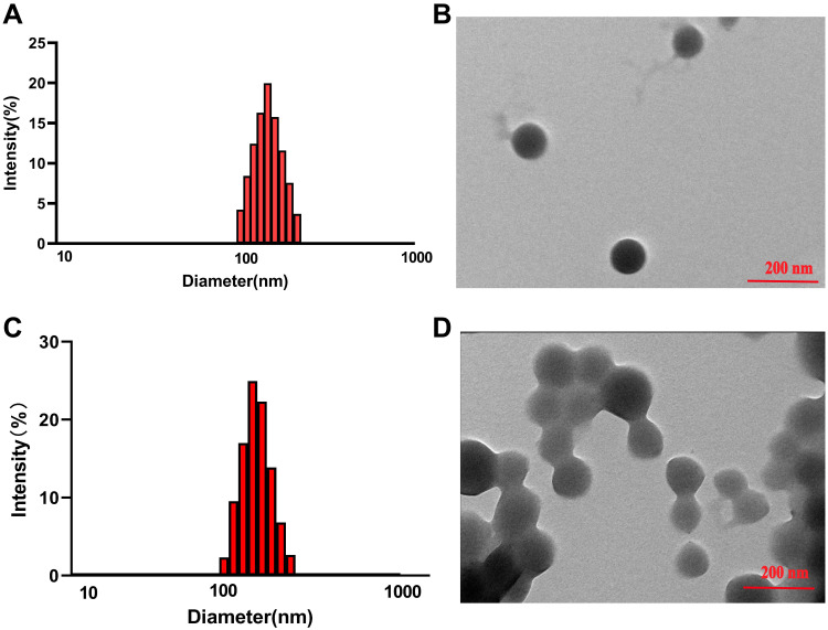
Maleimidocaproyl-valine-citrulline-p-aminobenzylcarbonyl-p-nitrophenol (MC-Val-Cit-PAB-PNP) and DOX were used to synthesize MC-Val-Cit-PAB-DOX, which was further linked with cetuximab to prepare antibody-drug conjugates (ADCs). Then, the ADCs were adsorbed to the surface of the BSA nanoparticles (NPs), which were prepared by a desolvation method to obtain cetuximab-vc-DOX-BSA-NPs. The cetuximab-vc-DOX conjugates adsorbed on the surface of the BSA nanoparticles were determined and optimized by size exclusion chromatography. An in vitro cytotoxicity study was conducted using a colon carcinoma cell line with different EGFR-expression levels to test the selectivity of cetuximab-vc-DOX-NPs.
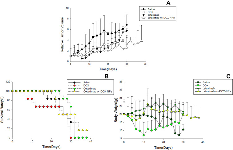
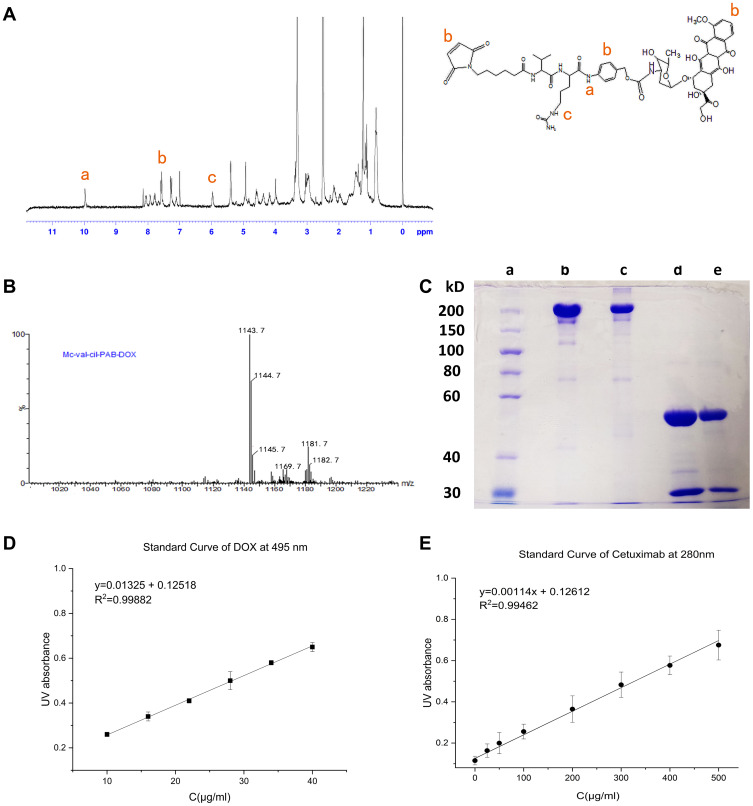
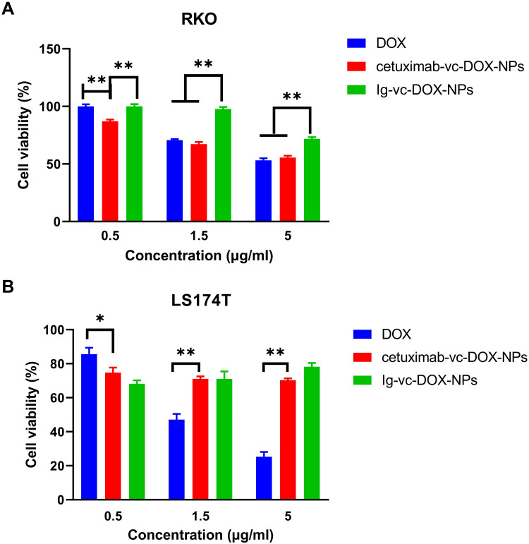
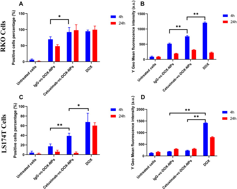
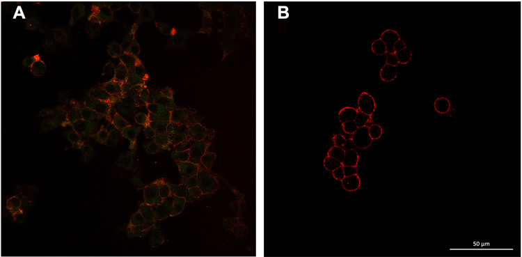
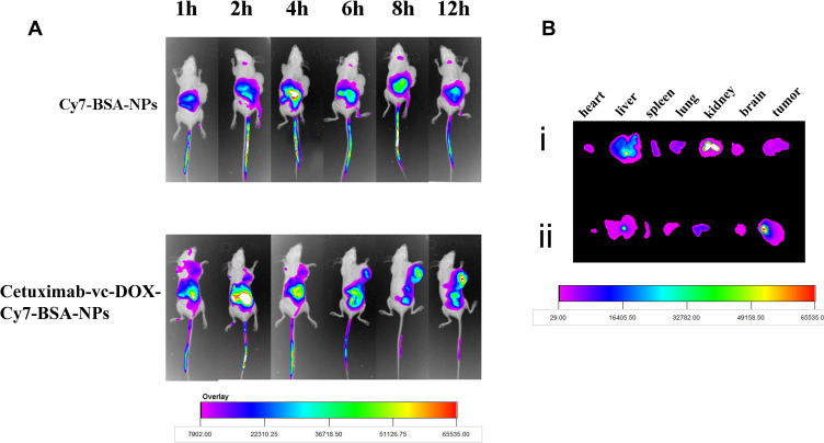
The vc-DOX and cetuximab-vc-DOX conjugates were both synthesized successfully and their structural characteristics confirmed by H-NMR and SDS-PAGE. The MTT assay showed stronger cytotoxicity of cetuximab-vc-DOX-NPs versus control IgG-vc-DOX-NPs in EGFR-overexpressing RKO cells. Cellular binding and intracellular accumulation determined by flow cytometry and confocal laser scanning microscopy revealed the strong binding ability of cetuximab-vc-DOX-NPs to RKO cells. The in vivo imaging study demonstrated that cetuximab-vc-DOX-NPs exhibited higher fluorescent intensity in tumor tissues than non-decorated nanoparticles (IgG-vc-DOX-NPs). In vivo tumor inhibition and survival tests showed that cetuximab-vc-DOX-NPs revealed higher tumor inhibition efficacy and lower systemic toxicity than control IgG-vc-DOX- NPs.
The obtained results emphasize that cetuximab-vc-DOX-NPs, with good tumor-targeting ability and low systemic toxicity, are a promising targeting system for drug delivery.
为了实现化疗药物对恶性组织的靶向递送,对载体进行特定修饰是提高诊断和治疗效率的关键。在本案例中,牛血清白蛋白(BSA)与西妥昔单抗-缬氨酸-瓜氨酸(vc)-阿霉素(DOX)偶联,以靶向表皮生长因子受体(EGFR),并使药物在 EGFR 过表达的肿瘤细胞中释放。
使用马来酰亚胺基己酰基-缬氨酸-瓜氨酸-p-氨基苯甲酰基-p-硝基苯酚(MC-Val-Cit-PAB-PNP)和 DOX 合成 MC-Val-Cit-PAB-DOX,进一步与西妥昔单抗偶联制备抗体药物偶联物(ADC)。然后,通过去溶剂法将 ADC 吸附到牛血清白蛋白纳米颗粒(NPs)表面,制备牛血清白蛋白-西妥昔单抗-vc-DOX 纳米颗粒(cetuximab-vc-DOX-BSA-NPs)。通过尺寸排阻色谱法确定和优化吸附在牛血清白蛋白纳米颗粒表面的西妥昔单抗-vc-DOX 缀合物。使用具有不同 EGFR 表达水平的结肠癌细胞系进行体外细胞毒性研究,以测试 cetuximab-vc-DOX-NPs 的选择性。
成功合成了 vc-DOX 和 cetuximab-vc-DOX 缀合物,并通过 H-NMR 和 SDS-PAGE 确认了它们的结构特征。MTT 测定表明,在 EGFR 过表达的 RKO 细胞中,cetuximab-vc-DOX-NPs 比对照 IgG-vc-DOX-NPs 具有更强的细胞毒性。通过流式细胞术和共聚焦激光扫描显微镜确定的细胞结合和细胞内积累表明,cetuximab-vc-DOX-NPs 与 RKO 细胞具有很强的结合能力。体内成像研究表明,cetuximab-vc-DOX-NPs 在肿瘤组织中的荧光强度高于未修饰的纳米颗粒(IgG-vc-DOX-NPs)。体内肿瘤抑制和生存试验表明,cetuximab-vc-DOX-NPs 比对照 IgG-vc-DOX-NPs 具有更高的肿瘤抑制疗效和更低的全身毒性。
研究结果强调了 cetuximab-vc-DOX-NPs 具有良好的肿瘤靶向能力和低全身毒性,是一种有前途的药物递送靶向系统。