Department of Rheumatology and Clinical Immunology, Peking Union Medical College Hospital, Chinese Academy of Medical Sciences & Peking Union Medical College, Beijing, China.
Key Laboratory of Rheumatology and Clinical Immunology, Ministry of Education, Beijing, China.
Front Immunol. 2021 Mar 19;12:578548. doi: 10.3389/fimmu.2021.578548. eCollection 2021.
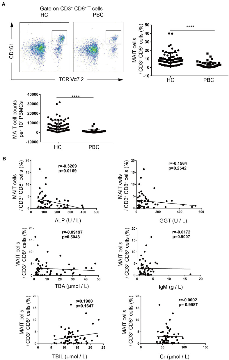
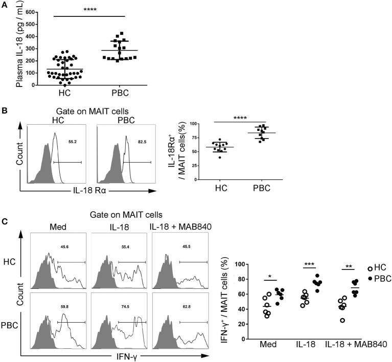
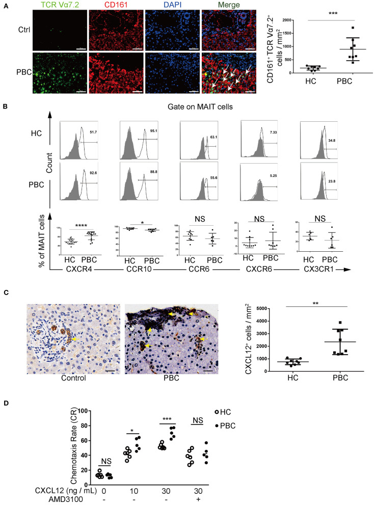
To explore the potential role of CD3CD8CD161 TCRVα7.2 mucosal-associated invariant T (MAIT) cells in the pathogenesis of primary biliary cholangitis (PBC). We enrolled 55 patients with PBC, 69 healthy controls (HCs), and 8 patients with hepatic hemangioma. Circulating MAIT cells and their chemokine receptor profiles and cytokine production were quantified using flow cytometry. Liver-resident MAIT cells were examined by immunofluorescence staining. CXCL12-mediated chemotaxis of MAIT cells was measured using a transwell migration assay. Plasma interleukin (IL)-18 was measured using ELISA, and cytokine production in IL-18-stimulated MAIT cells was detected using flow cytometry. Peripheral MAIT cells were found to be significantly lower in patients with PBC (3.0 ± 3.2% vs. 9.4 ± 8.0%, < 0.01) and negatively correlated with alkaline phosphatase (ALP) levels ( = -0.3209, < 0.05). Liver immunofluorescence staining suggested that MAIT cells might accumulate in PBC liver. MAIT cells from patients with PBC expressed higher levels of CXCR4 (84.8 ± 18.0% vs. 58.7 ± 11.4%, < 0.01), and the expression of CXCL12 was higher in PBC liver. CXCL12 promoted MAIT cell chemotaxis (70.4 ± 6.8% vs. 52.2 ± 3.5%, < 0.01), which was attenuated by CXCR4 antagonist. MAIT cells from PBC produced significantly more interferon-γ (IFN-γ) (88.3 ± 4.2% vs. 64.2 ± 10.1%, < 0.01), tumor necrosis factor-α (TNF-α) (93.0 ± 1.1% vs. 80.1 ± 5.3%, < 0.01), Granzyme B (89.3 ± 3.3% vs. 72.1 ± 7.0%, < 0.01), and perforin (46.8 ± 6.6% vs. 34.8 ± 7.7%, < 0.05). MAIT cells from PBC expressed higher levels of IL18-Rα (83.8 ± 10.2% vs. 58.3 ± 8.7%, < 0.01). Plasma IL-18 was more abundant in patients with PBC (286.8 ± 75.7 pg/ml vs. 132.9 ± 78.1 pg/ml, < 0.01). IL-18 promoted IFN-γ production in MAIT cells (74.9 ± 6.6% vs. 54.7 ± 6.7%, < 0.01), which was partially attenuated by blocking IL-18R (68.6 ± 8.3% vs. 43.5 ± 4.2%, < 0.01). Mucosal-associated invariant T cells from patients with PBC accumulated in the liver CXCL12-CXCR4-mediated chemotaxis, produced pro-inflammatory cytokines, and contributed to portal inflammation, which was potentially mediated by elevated IL-18. Targeting MAIT cells might be a therapeutic approach for PBC.
探讨 CD3CD8CD161 TCRVα7.2 黏膜相关不变 T(MAIT)细胞在原发性胆汁性胆管炎(PBC)发病机制中的潜在作用。我们纳入了 55 名 PBC 患者、69 名健康对照者(HCs)和 8 名肝血管瘤患者。使用流式细胞术定量检测循环 MAIT 细胞及其趋化因子受体谱和细胞因子产生。通过免疫荧光染色检查肝固有 MAIT 细胞。使用 Transwell 迁移测定法测量 CXCL12 介导的 MAIT 细胞趋化性。使用 ELISA 测量血浆白细胞介素(IL)-18,使用流式细胞术检测 IL-18 刺激的 MAIT 细胞中的细胞因子产生。发现 PBC 患者外周 MAIT 细胞明显降低(3.0±3.2%比 9.4±8.0%, <0.01),与碱性磷酸酶(ALP)水平呈负相关( = -0.3209, <0.05)。肝免疫荧光染色表明 MAIT 细胞可能在 PBC 肝脏中积聚。PBC 患者的 MAIT 细胞表达更高水平的 CXCR4(84.8±18.0%比 58.7±11.4%, <0.01),PBC 肝脏中 CXCL12 的表达更高。CXCL12 促进 MAIT 细胞趋化性(70.4±6.8%比 52.2±3.5%, <0.01),该趋化性被 CXCR4 拮抗剂减弱。PBC 的 MAIT 细胞产生显著更多的干扰素-γ(IFN-γ)(88.3±4.2%比 64.2±10.1%, <0.01)、肿瘤坏死因子-α(TNF-α)(93.0±1.1%比 80.1±5.3%, <0.01)、颗粒酶 B(89.3±3.3%比 72.1±7.0%, <0.01)和穿孔素(46.8±6.6%比 34.8±7.7%, <0.05)。PBC 的 MAIT 细胞表达更高水平的 IL18-Rα(83.8±10.2%比 58.3±8.7%, <0.01)。PBC 患者的血浆 IL-18 更丰富(286.8±75.7 pg/ml 比 132.9±78.1 pg/ml, <0.01)。IL-18 促进 MAIT 细胞 IFN-γ产生(74.9±6.6%比 54.7±6.7%, <0.01),该作用被阻断 IL-18R 部分减弱(68.6±8.3%比 43.5±4.2%, <0.01)。PBC 患者的黏膜相关不变 T 细胞在肝脏中积累 CXCL12-CXCR4 介导的趋化性,产生促炎细胞因子,并有助于门脉炎症,这可能是由升高的 IL-18 介导的。靶向 MAIT 细胞可能是治疗 PBC 的一种方法。