Asakawa Kazuhide, Handa Hiroshi, Kawakami Koichi
Department of Chemical Biology, Tokyo Medical University, Tokyo, Japan.
Division of Molecular and Developmental Biology, National Institute of Genetics, Mishima, Japan.
Front Cell Dev Biol. 2021 Mar 18;9:640414. doi: 10.3389/fcell.2021.640414. eCollection 2021.
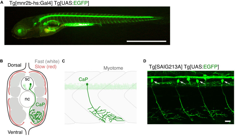
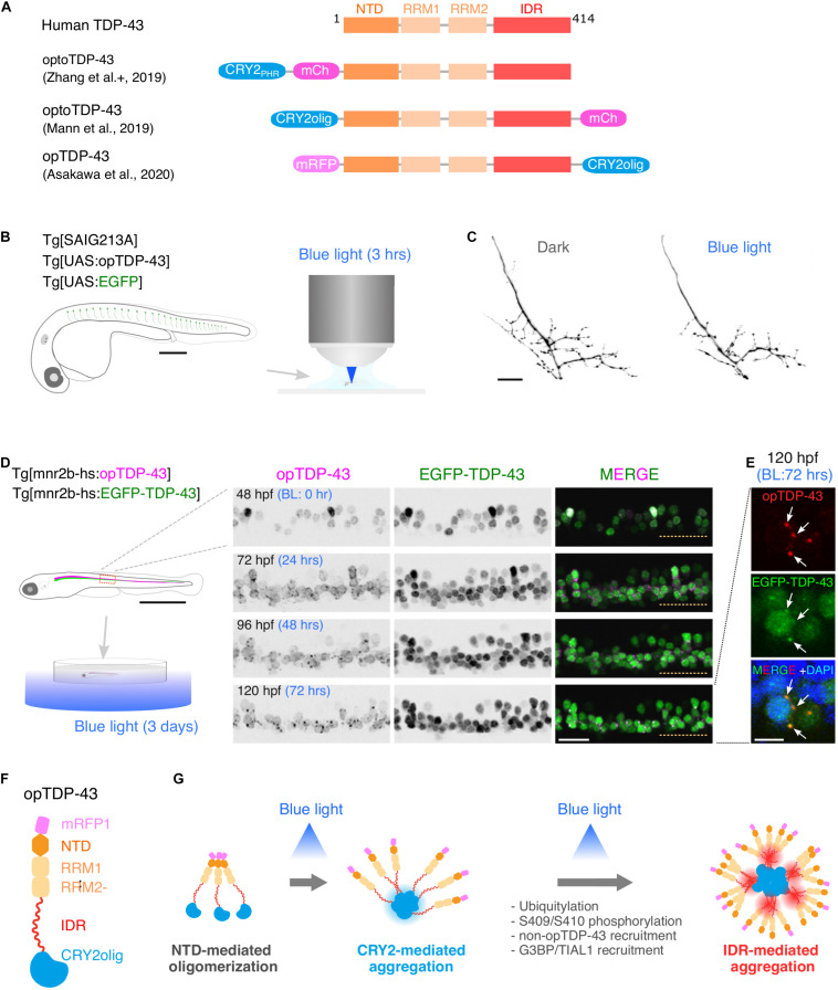
Amyotrophic lateral sclerosis (ALS) is a fatal neurological disorder characterized by progressive degeneration of motor neurons in the brain and spinal cord. Spinal motor neurons align along the spinal cord length within the vertebral column, and extend long axons to connect with skeletal muscles covering the body surface. Due to this anatomy, spinal motor neurons are among the most difficult cells to observe . Larval zebrafish have transparent bodies that allow non-invasive visualization of whole cells of single spinal motor neurons, from somas to the neuromuscular synapses. This unique feature, combined with its amenability to genome editing, pharmacology, and optogenetics, enables functional analyses of ALS-associated proteins in the spinal motor neurons with subcellular resolution. Here, we review the zebrafish skeletal neuromuscular system and the optical methods used to study it. We then introduce a recently developed optogenetic zebrafish ALS model that uses light illumination to control oligomerization, phase transition and aggregation of the ALS-associated DNA/RNA-binding protein called TDP-43. Finally, we will discuss how this disease-in-a-fish ALS model can help solve key questions about ALS pathogenesis and lead to new ALS therapeutics.
肌萎缩侧索硬化症(ALS)是一种致命的神经疾病,其特征是大脑和脊髓中的运动神经元进行性退化。脊髓运动神经元沿着脊柱内的脊髓长度排列,并延伸出长轴突以连接覆盖身体表面的骨骼肌。由于这种解剖结构,脊髓运动神经元是最难观察的细胞之一。斑马鱼幼体具有透明的身体,可对单个脊髓运动神经元的全细胞进行非侵入性可视化观察,从胞体到神经肌肉突触。这一独特特性,再加上其对基因组编辑、药理学和光遗传学的适应性,使得能够在亚细胞分辨率下对脊髓运动神经元中与ALS相关的蛋白质进行功能分析。在这里,我们综述斑马鱼骨骼神经肌肉系统以及用于研究它的光学方法。然后,我们介绍一种最近开发的光遗传学斑马鱼ALS模型,该模型利用光照来控制与ALS相关的DNA/RNA结合蛋白TDP-43的寡聚化、相变和聚集。最后,我们将讨论这种鱼类ALS疾病模型如何有助于解决有关ALS发病机制的关键问题,并催生新的ALS治疗方法。