• 文献检索
  • 文档翻译
  • 深度研究
  • 学术资讯
  • Suppr Zotero 插件Zotero 插件
  • 邀请有礼
  • 套餐&价格
  • 历史记录
应用&插件
Suppr Zotero 插件Zotero 插件浏览器插件Mac 客户端Windows 客户端微信小程序
定价
高级版会员购买积分包购买API积分包
服务
文献检索文档翻译深度研究API 文档MCP 服务
关于我们
关于 Suppr公司介绍联系我们用户协议隐私条款
关注我们

Suppr 超能文献

核心技术专利:CN118964589B侵权必究
粤ICP备2023148730 号-1Suppr @ 2026

文献检索

告别复杂PubMed语法,用中文像聊天一样搜索,搜遍4000万医学文献。AI智能推荐,让科研检索更轻松。

立即免费搜索

文件翻译

保留排版,准确专业,支持PDF/Word/PPT等文件格式,支持 12+语言互译。

免费翻译文档

深度研究

AI帮你快速写综述,25分钟生成高质量综述,智能提取关键信息,辅助科研写作。

立即免费体验

在使用多种CBCT设置的情况下,评估在存在3种不同牙科植入材料时,采集前与采集后金属伪影减少算法的效率:一项体外研究。

Assessment of the efficiency of a pre- versus post-acquisition metal artifact reduction algorithm in the presence of 3 different dental implant materials using multiple CBCT settings: An in vitro study.

作者信息

Shahmirzadi Solaleh, Sharaf Rana A, Saadat Sarang, Moore William S, Geha Hassem, Tamimi Dania, Demirturk Kocasarac Husniye

机构信息

Department of Diagnostic Sciences, Division of Oral and Maxillofacial Radiology, Texas A&M College of Dentistry, Dallas, TX, USA.

Department of Comprehensive Dentistry, Division of Oral and Maxillofacial Radiology, University of Texas Health Science Center, San Antonio, TX, USA.

出版信息

Imaging Sci Dent. 2021 Mar;51(1):1-7. doi: 10.5624/isd.20200094. Epub 2021 Jan 28.

DOI:10.5624/isd.20200094
PMID:33828955
原文链接:https://pmc.ncbi.nlm.nih.gov/articles/PMC8007401/
Abstract

PURPOSE

The aim of this study was to assess artifacts generated in cone-beam computed tomography (CBCT) of 3 types of dental implants using 3 metal artifact reduction (MAR) algorithm conditions (pre-acquisition MAR, postacquisition MAR, and no MAR), and 2 peak kilovoltage (kVp) settings.

MATERIALS AND METHODS

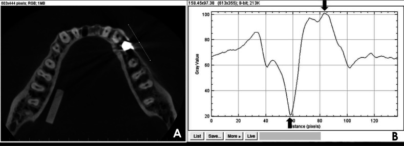
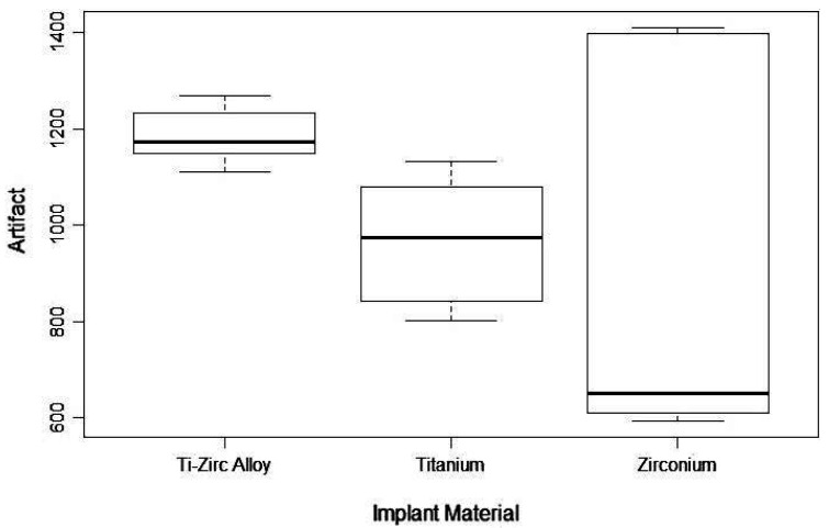
Titanium-zirconium, titanium, and zirconium alloy implants were placed in a dry mandible. CBCT images were acquired using 84 and 90 kVp and at normal resolution for all 3 MAR conditions. The images were analyzed using ImageJ software (National Institutes of Health, Bethesda, MD) to calculate the intensity of artifacts for each combination of material and settings. A 3-factor analysis of variance model with up to 3-way interactions was used to determine whether there was a statistically significant difference in the mean intensity of artifacts associated with each factor.

RESULTS

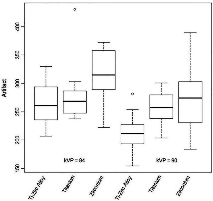
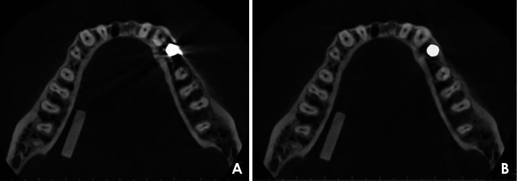
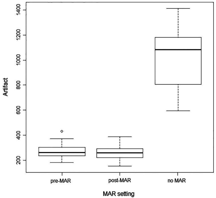
The analysis of all 3 MAR conditions showed that using no MAR resulted in substantially more severe artifacts than either of the 2 MAR algorithms for the 3 implant materials; however, there were no significant differences between pre- and post-acquisition MAR. The 90 kVp setting generated less intense artifacts on average than the 84 kVp setting. The titanium-zirconium alloy generated significantly less intense artifacts than zirconium. Titanium generated artifacts at an intermediate level relative to the other 2 implant materials, but was not statistically significantly different from either.

CONCLUSION

This in vitro study suggests that artifacts can be minimized by using a titanium-zirconium alloy at the 90 kVp setting, with either MAR setting.

摘要

目的

本研究旨在评估在三种金属伪影减少(MAR)算法条件(采集前MAR、采集后MAR和无MAR)以及两种峰值千伏(kVp)设置下,三种类型牙种植体的锥形束计算机断层扫描(CBCT)中产生的伪影。

材料与方法

将钛锆合金、钛和锆合金种植体植入干燥的下颌骨中。使用84和90 kVp并以正常分辨率采集所有三种MAR条件下的CBCT图像。使用ImageJ软件(美国国立卫生研究院,马里兰州贝塞斯达)对图像进行分析,以计算每种材料和设置组合的伪影强度。使用具有高达三向交互作用的三因素方差分析模型来确定与每个因素相关的伪影平均强度是否存在统计学上的显著差异。

结果

对所有三种MAR条件的分析表明,对于三种种植体材料,不使用MAR产生的伪影比两种MAR算法中的任何一种都要严重得多;然而,采集前和采集后MAR之间没有显著差异。90 kVp设置平均产生的伪影比84 kVp设置少。钛锆合金产生的伪影强度明显低于锆。钛产生的伪影相对于其他两种种植体材料处于中等水平,但与两者均无统计学上的显著差异。

结论

这项体外研究表明,在90 kVp设置下,使用钛锆合金并结合任何一种MAR设置,可将伪影最小化。

https://cdn.ncbi.nlm.nih.gov/pmc/blobs/4a60/8007401/17ecc153abfa/isd-51-1-g005.jpg
https://cdn.ncbi.nlm.nih.gov/pmc/blobs/4a60/8007401/2bb1a6fb5eb0/isd-51-1-g001.jpg
https://cdn.ncbi.nlm.nih.gov/pmc/blobs/4a60/8007401/7ee49ff9e933/isd-51-1-g002.jpg
https://cdn.ncbi.nlm.nih.gov/pmc/blobs/4a60/8007401/21b196556aa4/isd-51-1-g003.jpg
https://cdn.ncbi.nlm.nih.gov/pmc/blobs/4a60/8007401/a82815346b8a/isd-51-1-g004.jpg
https://cdn.ncbi.nlm.nih.gov/pmc/blobs/4a60/8007401/17ecc153abfa/isd-51-1-g005.jpg

相似文献

1
Assessment of the efficiency of a pre- versus post-acquisition metal artifact reduction algorithm in the presence of 3 different dental implant materials using multiple CBCT settings: An in vitro study.在使用多种CBCT设置的情况下,评估在存在3种不同牙科植入材料时,采集前与采集后金属伪影减少算法的效率:一项体外研究。
Imaging Sci Dent. 2021 Mar;51(1):1-7. doi: 10.5624/isd.20200094. Epub 2021 Jan 28.
2
Evaluation of artifacts generated by zirconium implants in cone-beam computed tomography images.锥形束计算机断层扫描图像中锆植入物产生的伪影评估。
Oral Surg Oral Med Oral Pathol Oral Radiol. 2017 Feb;123(2):265-272. doi: 10.1016/j.oooo.2016.10.021. Epub 2016 Nov 1.
3
Influence of Cone Beam Computed Tomography Settings on Implant Artifact Production: Zirconia and Titanium.锥形束计算机断层扫描设置对种植体伪影产生的影响:氧化锆和钛。
Int J Oral Maxillofac Implants. 2019 Sep/Oct;34(5):1114-1120. doi: 10.11607/jomi.7129.
4
Influence of artefacts generated by titanium and zirconium implants in the study of trabecular bone architecture in cone-beam CT images.钛和锆植入物产生的伪影对锥形束 CT 图像中小梁骨结构研究的影响。
Dentomaxillofac Radiol. 2022 Sep 1;51(6):20220066. doi: 10.1259/dmfr.20220066. Epub 2022 May 4.
5
Comparative Evaluation of Artifacts Originated by Four Different Post Materials Using Different CBCT Settings.四种不同根管材料在不同 CBCT 设置下产生的伪影的比较评估。
Tomography. 2022 Dec 13;8(6):2919-2928. doi: 10.3390/tomography8060245.
6
Do the tube current and metal artifact reduction influence the diagnosis of vertical root fracture in a tooth positioned in the vicinity of a zirconium implant? A CBCT study.管电流和金属伪影减少技术是否会影响临近氧化锆种植体的牙的垂直根折诊断?一项 CBCT 研究。
Clin Oral Investig. 2021 Apr;25(4):2229-2235. doi: 10.1007/s00784-020-03538-4. Epub 2020 Aug 22.
7
Application of an auto-edge counting method for quantification of metal artifacts in CBCT images: a multivariate analysis of object position, field of view size, tube voltage, and metal artifact reduction algorithm.应用自动边缘计数法对锥形束 CT 图像中的金属伪影进行定量分析:物体位置、视野大小、管电压和金属伪影降低算法的多变量分析。
Oral Surg Oral Med Oral Pathol Oral Radiol. 2021 Dec;132(6):735-743. doi: 10.1016/j.oooo.2021.03.012. Epub 2021 Mar 28.
8
Diagnosis of vertical root fracture in teeth close and distant to implant: an in vitro study to assess the influence of artifacts produced in cone beam computed tomography.诊断近中、远中种植体邻牙的垂直根折:评估锥形束计算机断层扫描产生的伪影影响的体外研究。
Clin Oral Investig. 2019 Mar;23(3):1263-1270. doi: 10.1007/s00784-018-2558-z. Epub 2018 Jul 8.
9
Does the metal artifact reduction algorithm activation mode influence the magnitude of artifacts in CBCT images?金属伪影减少算法的激活模式会影响锥形束计算机断层扫描(CBCT)图像中的伪影大小吗?
Imaging Sci Dent. 2020 Mar;50(1):23-30. doi: 10.5624/isd.2020.50.1.23. Epub 2020 Mar 17.
10
CBCT image artefacts generated by implants located inside the field of view or in the exomass.视野内或外源性物质内的植入物产生的 CBCT 图像伪影。
Dentomaxillofac Radiol. 2022 Feb 1;51(2):20210092. doi: 10.1259/dmfr.20210092. Epub 2021 Jul 29.

引用本文的文献

1
Quantitative evaluation of metal artifact reduction in CBCT under varying exposure modes and rod orientations.在不同曝光模式和棒状物体取向条件下,对锥形束CT(CBCT)中金属伪影减少情况的定量评估。
Sci Rep. 2025 Jul 1;15(1):20645. doi: 10.1038/s41598-025-08188-8.
2
Efficacy of two radiographic algorithms for detection of peri-implant bone defects on cone-beam computed tomography scans.两种影像学算法在锥形束计算机断层扫描上检测种植体周围骨缺损的效能
BMC Oral Health. 2025 Jan 7;25(1):39. doi: 10.1186/s12903-024-05397-x.
3
Evaluation of clinical and radiographic warning signs for prediction of oroantral communication following tooth extractions.

本文引用的文献

1
Evaluation of artifacts generated by titanium, zirconium, and titanium-zirconium alloy dental implants on MRI, CT, and CBCT images: A phantom study.评价钛、锆和钛锆合金牙种植体在 MRI、CT 和 CBCT 图像上产生的伪影:一项体模研究。
Oral Surg Oral Med Oral Pathol Oral Radiol. 2019 Jun;127(6):535-544. doi: 10.1016/j.oooo.2019.01.074. Epub 2019 Feb 7.
2
Evaluation of artifacts generated by zirconium implants in cone-beam computed tomography images.锥形束计算机断层扫描图像中锆植入物产生的伪影评估。
Oral Surg Oral Med Oral Pathol Oral Radiol. 2017 Feb;123(2):265-272. doi: 10.1016/j.oooo.2016.10.021. Epub 2016 Nov 1.
3
评估临床和影像学警告标志以预测拔牙后发生上颌窦-口腔交通。
Clin Oral Investig. 2024 Oct 23;28(11):609. doi: 10.1007/s00784-024-06000-x.
4
Influence of Windowing and Evaluation of Metal Artifact Reduction Algorithm on Five Different Restorative Materials by Using Different Cone Beam Computed Tomography (CBCT) Scanners: A CBCT Study.使用不同锥形束计算机断层扫描(CBCT)扫描仪对五种不同修复材料进行开窗及金属伪影减少算法评估的影响:一项CBCT研究
Cureus. 2023 Jul 11;15(7):e41742. doi: 10.7759/cureus.41742. eCollection 2023 Jul.
5
The Effect of Changes in the Angular Position of Implants on Metal Artifact Reduction in Cone-Beam Computed Tomography Images: A Scoping Review.种植体角度位置变化对锥形束计算机断层扫描图像中金属伪影减少的影响:一项范围综述
Radiol Res Pract. 2023 Jul 31;2023:5539719. doi: 10.1155/2023/5539719. eCollection 2023.
6
Efficacy of low-dose cone beam computed tomography and metal artifact reduction tool for assessment of peri-implant bone defects: an in vitro study.低剂量锥形束 CT 和金属伪影降低工具评估种植体周围骨缺损的疗效:一项体外研究。
BMC Oral Health. 2022 Dec 17;22(1):615. doi: 10.1186/s12903-022-02663-8.
7
Comparison of the amount of artifacts induced by zirconium and titanium implants in cone-beam computed tomography images.比较锥形束计算机断层摄影图像中锆和钛植入物引起的伪影量。
BMC Med Imaging. 2022 Sep 3;22(1):156. doi: 10.1186/s12880-022-00884-5.
8
Quantitative Evaluation of Postprocessing Filter for Metal Artifact Reduction in Cone Beam Computed Tomography Images of Titanium and Zirconium Dioxide Implants.钛和二氧化锆种植体锥形束计算机断层摄影图像中金属伪影减少的后处理滤波器的定量评估。
Biomed Res Int. 2022 Feb 24;2022:1362473. doi: 10.1155/2022/1362473. eCollection 2022.
A systematic review of the clinical survival of zirconia implants.
氧化锆种植体临床生存率的系统评价。
Clin Oral Investig. 2016 Sep;20(7):1403-17. doi: 10.1007/s00784-016-1853-9. Epub 2016 May 24.
4
Contrast-to-noise ratio with different settings in a CBCT machine in presence of different root-end filling materials: an in vitro study.在存在不同根尖充填材料的情况下,CBCT 机不同设置下的对比噪声比:一项体外研究。
Dentomaxillofac Radiol. 2016;45(5):20160012. doi: 10.1259/dmfr.20160012. Epub 2016 Mar 8.
5
Clinical evidence on titanium-zirconium dental implants: a systematic review and meta-analysis.钛锆牙种植体的临床证据:一项系统评价与荟萃分析。
Int J Oral Maxillofac Surg. 2016 Jul;45(7):842-50. doi: 10.1016/j.ijom.2016.01.004. Epub 2016 Feb 3.
6
Influence of the artefact reduction algorithm of Picasso Trio CBCT system on the diagnosis of vertical root fractures in teeth with metal posts.毕加索三重CBCT系统的伪影减少算法对带有金属桩牙齿垂直根折诊断的影响
Dentomaxillofac Radiol. 2015;44(6):20140428. doi: 10.1259/dmfr.20140428. Epub 2015 Mar 12.
7
Assessment of metal artefact reduction around dental titanium implants in cone beam CT.锥形束CT中牙科钛种植体周围金属伪影减少的评估
Dentomaxillofac Radiol. 2014;43(7):20140019. doi: 10.1259/dmfr.20140019. Epub 2014 Aug 19.
8
In vitro assessment of artifacts induced by titanium, titanium-zirconium and zirconium dioxide implants in cone-beam computed tomography.锥形束计算机断层扫描中钛、钛锆和二氧化锆种植体引起的伪影的体外评估。
Clin Oral Implants Res. 2015 Oct;26(10):1222-8. doi: 10.1111/clr.12438. Epub 2014 Jul 8.
9
Accuracy of CBCT images in the assessment of buccal marginal alveolar peri-implant defects: effect of field of view.CBCT 图像评估颊侧边缘牙槽嵴种植体周围缺损的准确性:视野的影响。
Dentomaxillofac Radiol. 2014;43(4):20130332. doi: 10.1259/dmfr.20130332. Epub 2014 Mar 20.
10
Assessment of buccal marginal alveolar peri-implant and periodontal defects using a cone beam CT system with and without the application of metal artefact reduction mode.使用配备和不配备金属伪影降低模式的锥形束 CT 系统评估颊侧边缘牙槽嵴种植体和牙周缺损。
Dentomaxillofac Radiol. 2013;42(8):20130176. doi: 10.1259/dmfr.20130176.