Department of Biomedical Engineering, Duke University, Durham, North Carolina, USA.
Department of Biomedical Engineering, Duke University, Durham, North Carolina, USA.
Ultrasound Med Biol. 2021 Jul;47(7):1670-1680. doi: 10.1016/j.ultrasmedbio.2021.02.006. Epub 2021 Apr 6.
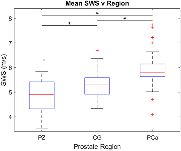
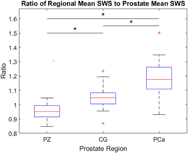
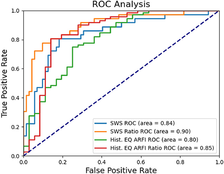
Transrectal ultrasound (TRUS) B-mode imaging provides insufficient sensitivity and specificity for prostate cancer (PCa) targeting when used for biopsy guidance. Shear wave elasticity imaging (SWEI) is an elasticity imaging technique that has been commercially implemented and is sensitive and specific for PCa. We have developed a SWEI system capable of 3-D data acquisition using a dense acoustic radiation force (ARF) push approach that leads to enhanced shear wave signal-to-noise ratio compared with that of the commercially available SWEI systems and facilitates screening of the entire gland before biopsy. Additionally, we imaged and assessed 36 patients undergoing radical prostatectomy using 3-D SWEI and determined a shear wave speed threshold separating PCa from healthy prostate tissue with sensitivities and specificities akin to those for multiparametric magnetic resonance imaging fusion biopsy. The approach measured the mean shear wave speed in each prostate region to be 4.8 m/s (Young's modulus E = 69.1 kPa) in the peripheral zone, 5.3 m/s (E = 84.3 kPa) in the central gland and 6.0 m/s (E = 108.0 kPa) for PCa with statistically significant (p < 0.0001) differences among all regions. Three-dimensional SWEI receiver operating characteristic analyses identified a threshold of 5.6 m/s (E = 94.1 kPa) to separate PCa from healthy tissue with a sensitivity, specificity, positive predictive value (PPV), negative predictive value (NPV) and area under the curve (AUC) of 81%, 82%, 69%, 89% and 0.84, respectively. Additionally, a shear wave speed ratio was assessed to normalize for tissue compression and patient variability, which yielded a threshold of 1.11 to separate PCa from healthy prostate tissue and was accompanied by a substantial increase in specificity, PPV and AUC, where the sensitivity, specificity, PPV, NPV and AUC were 75%, 90%, 79%, 88% and 0.90, respectively. This work illustrates the feasibility of using 3-D SWEI data to detect and localize PCa and demonstrates the benefits of normalizing for applied compression during data acquisition for use in biopsy targeting studies.
经直肠超声(TRUS)B 模式成像在用于活检引导时,对前列腺癌(PCa)的靶向定位的灵敏度和特异性不足。剪切波弹性成像(SWEI)是一种弹性成像技术,已商业化实施,对 PCa 具有敏感性和特异性。我们开发了一种 SWEI 系统,能够使用密集声辐射力(ARF)推送方法进行 3D 数据采集,与商业上可用的 SWEI 系统相比,这种方法可提高剪切波的信噪比,并在活检前方便地对整个腺体进行筛查。此外,我们使用 3D SWEI 对 36 例接受根治性前列腺切除术的患者进行了成像和评估,并确定了一个剪切波速度阈值,可将 PCa 与健康前列腺组织区分开来,其灵敏度和特异性与多参数磁共振成像融合活检相当。该方法测量每个前列腺区域的平均剪切波速度,在周边区为 4.8 m/s(杨氏模量 E=69.1 kPa),在中央区为 5.3 m/s(E=84.3 kPa),PCa 为 6.0 m/s(E=108.0 kPa),所有区域之间的差异均具有统计学意义(p<0.0001)。3D SWEI 的接收器操作特性分析确定了一个阈值为 5.6 m/s(E=94.1 kPa),可将 PCa 与健康组织分开,其灵敏度、特异性、阳性预测值(PPV)、阴性预测值(NPV)和曲线下面积(AUC)分别为 81%、82%、69%、89%和 0.84。此外,还评估了剪切波速度比以对组织压缩和患者变异性进行归一化,得到的阈值为 1.11,可将 PCa 与健康前列腺组织分开,并且特异性、PPV 和 AUC 显著增加,其灵敏度、特异性、PPV、NPV 和 AUC 分别为 75%、90%、79%、88%和 0.90。这项工作说明了使用 3D SWEI 数据检测和定位 PCa 的可行性,并证明了在数据采集过程中对施加的压缩进行归一化以用于活检靶向研究的益处。