IBFM-CNR, Segrate, Italy.
Experimental Imaging Center, IRCCS San Raffaele Scientific Institute, Milan, Italy.
J Neuroinflammation. 2018 Feb 5;15(1):33. doi: 10.1186/s12974-017-1044-x.
Positron emission tomography (PET) using translocator protein (TSPO) ligands has been used to detect neuroinflammatory processes in neurological disorders, including multiple sclerosis (MS). The aim of this study was to evaluate neuroinflammation in a mouse MS model (EAE) using TSPO-PET with F-VC701, in combination with magnetic resonance imaging (MRI).
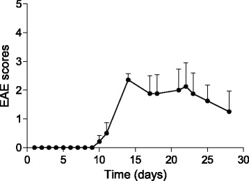
MOG/CFA and pertussis toxin protocol was used to induce EAE in C57BL/6 mice. Disease progression was monitored daily, whereas MRI evaluation was performed at 1, 2, and 4 weeks post-induction. Microglia activation was assessed in vivo by F-VC701 PET at the time of maximum disease score and validated by radioligand ex vivo distribution and immunohistochemistry at 2 and 4 weeks post-immunization.
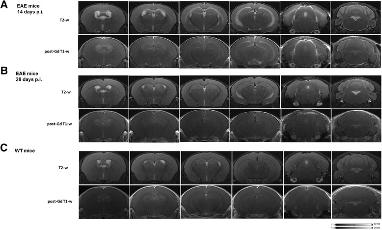
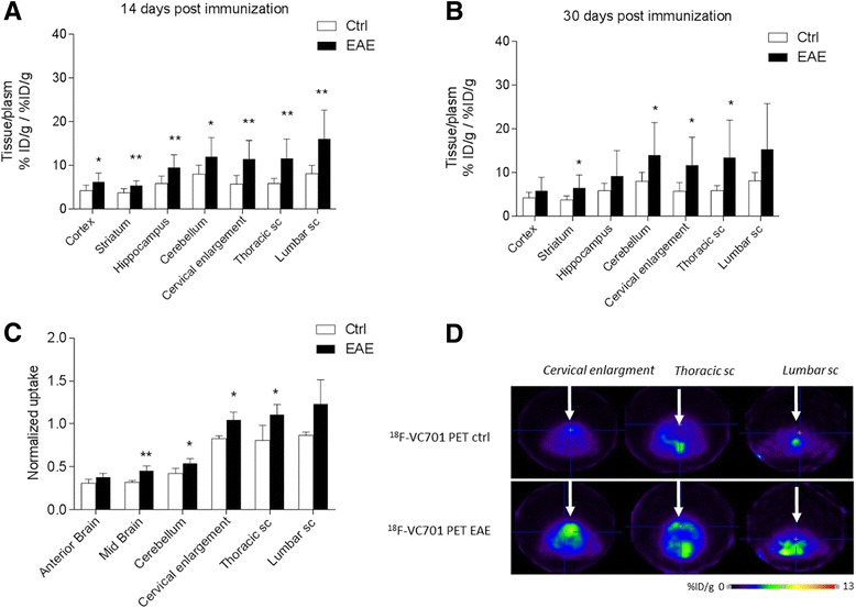
In vivo and ex vivo analyses show that F-VC701 significantly accumulates within the central nervous system (CNS), particularly in the cortex, striatum, hippocampus, cerebellum, and cervical spinal cord of EAE compared to control mice, at 2 weeks post-immunization. MRI confirmed the presence of focal brain lesions at 2 weeks post-immunization in both T1-weighted and T2 images. Of note, MRI abnormalities attenuated in later post-immunization phase. Neuropathological analysis confirmed the presence of microglial activation in EAE mice, consistent with the in vivo increase of F-VC701 uptake.
Increase of F-VC701 uptake in EAE mice is strongly associated with the presence of microglia activation in the acute phase of the disease. The combined use of TSPO-PET and MRI provided complementary evidence on the ongoing disease process, thus representing an attractive new tool to investigate neuronal damage and neuroinflammation at preclinical levels.
正电子发射断层扫描(PET)使用转位蛋白(TSPO)配体已被用于检测神经炎症过程在神经疾病,包括多发性硬化症(MS)。本研究的目的是评估 TSPO-PET 与 F-VC701 联合磁共振成像(MRI)在 MS 模型(EAE)中的神经炎症。
MOG/CFA 和百日咳毒素方案用于诱导 C57BL/6 小鼠 EAE。疾病进展情况每天监测,而 MRI 评估在诱导后 1、2 和 4 周进行。在疾病评分最高时通过 F-VC701 PET 评估体内小胶质细胞激活,并在免疫后 2 和 4 周通过放射性配体体外分布和免疫组织化学验证。
体内和体外分析表明,与对照组相比,F-VC701 在 EAE 小鼠的中枢神经系统(CNS)中特别是在大脑皮层、纹状体、海马、小脑和颈脊髓中显著积聚,在免疫后 2 周时。MRI 证实免疫后 2 周时 T1 加权和 T2 图像存在局灶性脑损伤。值得注意的是,MRI 异常在免疫后后期减轻。神经病理学分析证实 EAE 小鼠存在小胶质细胞激活,与 F-VC701 摄取的增加一致。
EAE 小鼠 F-VC701 摄取的增加与疾病急性期小胶质细胞激活的存在密切相关。TSPO-PET 和 MRI 的联合使用提供了关于疾病进展的互补证据,因此代表了一种有吸引力的新工具,可在临床前水平研究神经元损伤和神经炎症。