Department of Biochemistry, German University in Cairo, Egypt.
Institute of Biochemistry, University of Erlangen-Nürnberg, Germany.
J Adv Res. 2020 Oct 8;29:95-106. doi: 10.1016/j.jare.2020.09.009. eCollection 2021 Mar.
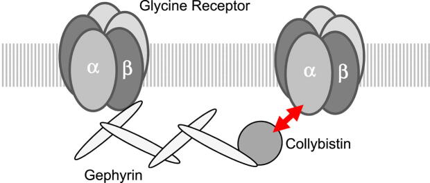
The inhibitory glycine receptor (GlyR), a mediator of fast synaptic inhibition, is located and held at neuronal synapses through the anchoring proteins gephyrin and collybistin. Stable localization of neurotransmitter receptors is essential for synaptic function. In case of GlyRs, only beta subunits were known until now to mediate synaptic anchoring.
We identified a poly-proline II helix (PPII) in position 365-373 of the intra-cellular TM3-4 loop of the human GlyRα1 subunit as a novel potential synaptic anchoring site. The potential role of the PPII helix as synaptic anchoring site was tested.
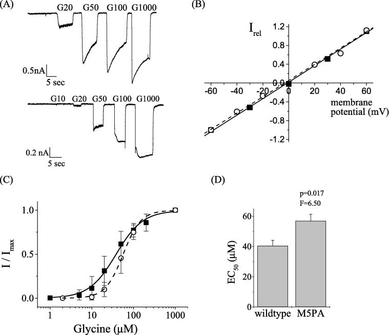
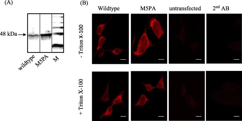
Glycine receptors and collybistin variants were generated and recombinantly expressed in HEK293 cells and cultured neurons. Receptor function was assessed using patch-clamp electrophysiology, protein-protein interaction was studied using co-immuno-precipitation and pulldown experiments.
Recombinantly expressed collybistin bound to isolated GlyRα1 TM3-4 loops in GST-pulldown assays. When the five proline residues P365A, P366A, P367A, P369A, P373A (GlyRα1) located in the GlyRα1-PPII helix were replaced by alanines, the PPII secondary structure was disrupted. Recombinant GlyRα1 mutant subunits displayed normal cell surface expression and wildtype-like ion channel function, but binding to collybistin was abolished. The GlyRα1-collybistin interaction was independently confirmed by o-immunoprecipitation assays using full-length GlyRα1 subunits. Surprisingly, the interaction was not mediated by the SH3 domain of collybistin, but by its Pleckstrin homology (PH) domain. The mutation GlyRα1, identified in a hyperekplexia patient, is also disrupting the PPII helix, and caused reduced collybistin binding.
Our data suggest a novel interaction between α1 GlyR subunits and collybistin, which is physiologically relevant in vitro and in vivo and may contribute to postsynaptic anchoring of glycine receptors.
抑制性甘氨酸受体(GlyR)是一种介导快速突触抑制的递质受体,通过锚定蛋白神经胶质蛋白和Collipstin 定位并保持在神经元突触上。神经递质受体的稳定定位对于突触功能至关重要。在 GlyR 的情况下,直到现在才知道只有β亚基介导突触锚定。
我们在人类 GlyRα1 亚基的细胞内 TM3-4 环的位置 365-373 处鉴定出一个多脯氨酸 II 螺旋(PPII)作为新的潜在突触锚定位点。测试了 PPII 螺旋作为突触锚定位点的潜在作用。
生成并在 HEK293 细胞和培养神经元中重组表达甘氨酸受体和 Collipstin 变体。使用膜片钳电生理学评估受体功能,使用共免疫沉淀和拉下实验研究蛋白-蛋白相互作用。
重组表达的 Collipstin 在 GST 下拉测定中与分离的 GlyRα1 TM3-4 环结合。当位于 GlyRα1-PPII 螺旋中的五个脯氨酸残基 P365A、P366A、P367A、P369A、P373A(GlyRα1)被替换为丙氨酸时,PPII 二级结构被破坏。重组 GlyRα1 突变亚基显示正常的细胞表面表达和野生型样离子通道功能,但与 Collipstin 的结合被废除。通过使用全长 GlyRα1 亚基的 o-免疫沉淀测定独立证实了 GlyRα1-Colliptin 相互作用。令人惊讶的是,这种相互作用不是由 Collipstin 的 SH3 结构域介导的,而是由其 Pleckstrin 同源(PH)结构域介导的。在一名发作性强肌阵挛性癫痫患者中发现的突变 GlyRα1 也破坏了 PPII 螺旋,并导致 Collipstin 结合减少。
我们的数据表明α1 GlyR 亚基和 Collipstin 之间存在新的相互作用,这种相互作用在体外和体内具有生理相关性,可能有助于甘氨酸受体的突触后锚定。