Immunobiology Department, Hospital Universitario Gregorio Marañon, Instituto de Investigación Sanitaria Gregorio Marañón (IisGM).
Department of Paediatric Infectious Diseases, Hospital Universitario 12 de Octubre; Instituto de Investigación Sanitaria Hospital 12 de Octubre (i+12).
Medicine (Baltimore). 2021 Apr 16;100(15):e25403. doi: 10.1097/MD.0000000000025403.
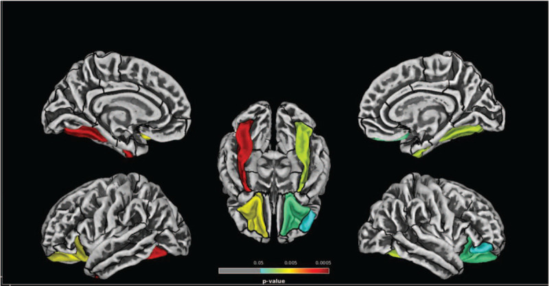
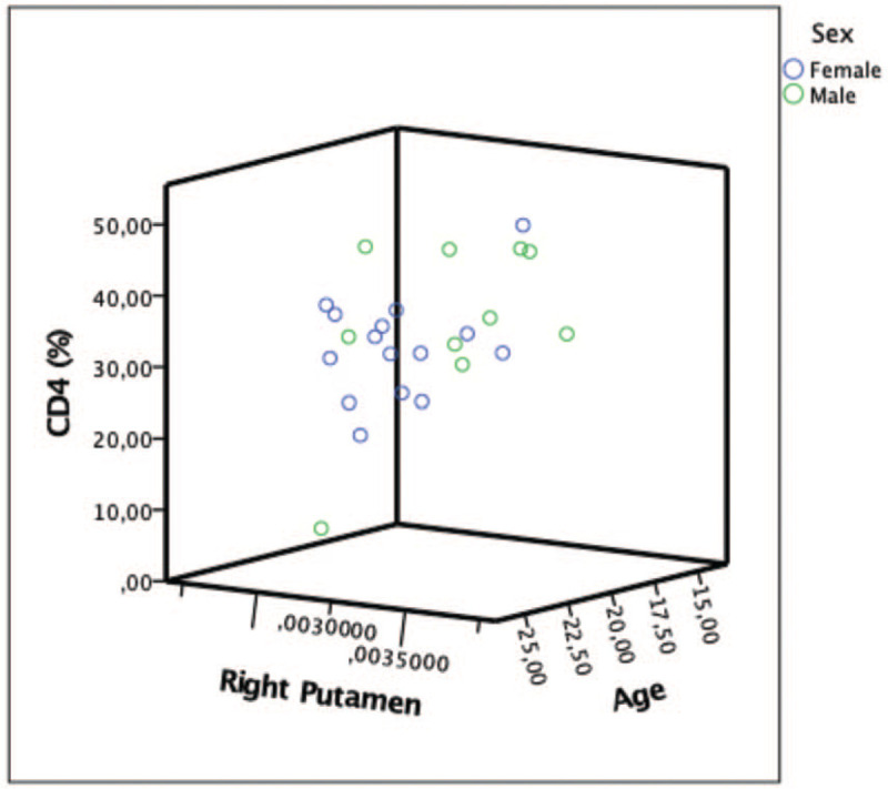
Brain atrophy has been observed in perinatally HIV-infected patients (PHIV) despite initiation on combined antiretroviral treatment (cART), but neuroimaging studies are limited. We aimed to evaluate cortical thickness (CT) and subcortical gray matter (GM) volumes of PHIV youths with stable immunovirological situation and with a normal daily performance.A prospective cross-sectional study was conducted. A total of 25 PHIV patients on cART and 25 HIV-negative (HIV-) controls matched by age, sex, level of education, and socioeconomic status underwent a magnetic resonance imaging scan. CAT12 toolbox was used to extract CT values from T1w images using parcellations from Desikan-Killiany atlas (DK40). To measure regional brain volumes, native segmented images were parceled in regions of interest according to the Neuromorphometrics Atlas. Neuropsychological assessment and psychopathological symptoms were documented.Fifty participants were included (60% females, median age 20 years [interquartile range, IQR 19-23], 64% Whites). No differences regarding neuropsychological tests or psychopathological symptoms were found between groups (all P > .05). All participants presented an average performance in the Fluid Intelligence (FI) test (PHIV mean: -0.12, HIV- mean: 0.24), When comparing CT, PHIV-infected patients showed thinner cortices compared with their peers in fusiform gyrus (P = .000, P = .009), lateral-orbitofrontal gyrus (P = .006, P = .0024), and right parsobitalis gyrus (P = .047). Regarding subcortical GM volumes, PHIV patients showed lower right amygdala (P = .014) and left putamen (P = .016) volumes when compared with HIV- controls. Within the PHIV group, higher CD4 count was associated with higher volumes in right putamen (B = 0.00000038, P = .045). Moreover, increased age at cART initiation and lower nadir CD4 count was associated with larger volumes in left accumbens (B = 0.0000046, P = .033; B = -0.00000008, P = .045, respectively).PHIV patients showed thinner cortices of areas in temporal, orbito-frontal and occipital lobes and lower volumes of subcortical GM volumes when compared with the HIV- control group, suggesting cortical and subcortical brain alterations in otherwise neuroasymptomatic patients. Nevertheless, larger and longitudinal studies are required to determine the impact of HIV on brain structure in PHIV patients and to further identify risk and protective factors that could be implicated.
脑萎缩在围生期 HIV 感染患者(PHIV)中是观察到的,尽管已经开始接受联合抗逆转录病毒治疗(cART),但神经影像学研究有限。我们旨在评估免疫病毒学稳定且日常表现正常的 PHIV 青少年的皮质厚度(CT)和皮质下灰质(GM)体积。
进行了一项前瞻性的横断面研究。共有 25 名接受 cART 的 PHIV 患者和 25 名 HIV 阴性(HIV-)对照者按年龄、性别、教育程度和社会经济地位进行匹配,接受了磁共振成像扫描。使用 Desikan-Killiany 图谱(DK40)的分区从 T1w 图像中提取 CT 值,使用 CAT12 工具箱。为了测量区域脑体积,将原始分割图像根据神经形态学图谱分区到感兴趣的区域。记录了神经心理学评估和精神病理学症状。
共有 50 名参与者(60%为女性,中位年龄 20 岁[四分位数范围 IQR 19-23],64%为白人)。两组之间在神经心理学测试或精神病理学症状方面没有差异(均 P>.05)。所有参与者在流体智力(FI)测试中的表现均平均(PHIV 平均值:-0.12,HIV-平均值:0.24)。比较 CT 时,PHIV 感染者的梭状回(P=0.000,P=0.009)、外侧额眶回(P=0.006,P=0.0024)和右侧旁矢状回(P=0.047)的皮质较薄。关于皮质下 GM 体积,与 HIV-对照组相比,PHIV 患者的右侧杏仁核(P=0.014)和左侧壳核(P=0.016)体积较低。在 PHIV 组中,较高的 CD4 计数与右侧壳核体积较大有关(B=0.00000038,P=0.045)。此外,cART 起始时年龄较大和较低的 CD4 计数最低点与左侧伏隔核体积较大有关(B=0.0000046,P=0.033;B=-0.00000008,P=0.045)。
与 HIV-对照组相比,PHIV 患者的颞叶、眶额和枕叶区域的皮质较薄,皮质下 GM 体积较低,提示神经无症状患者存在皮质和皮质下脑改变。然而,需要更大和纵向的研究来确定 HIV 对 PHIV 患者大脑结构的影响,并进一步确定可能涉及的风险和保护因素。