Department of Neurodegenerative Disease, Institute of Neurology, UCL, London, United Kingdom.
Department of Computer Science and Centre for Medical Image Computing, UCL, London, United Kingdom.
Hum Brain Mapp. 2018 Jul;39(7):3005-3017. doi: 10.1002/hbm.24056. Epub 2018 Mar 25.
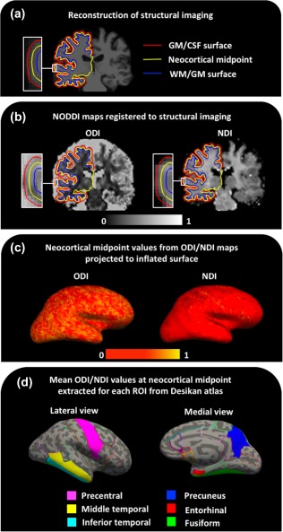
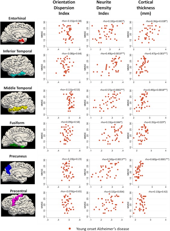
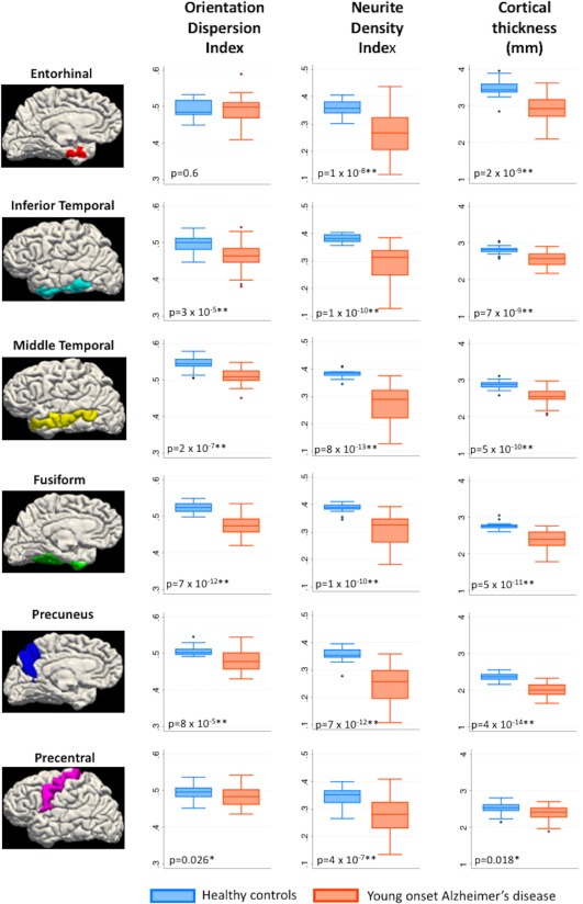
Alzheimer's disease (AD) is associated with extensive alterations in grey matter microstructure, but our ability to quantify this in vivo is limited. Neurite orientation dispersion and density imaging (NODDI) is a multi-shell diffusion MRI technique that estimates neuritic microstructure in the form of orientation dispersion and neurite density indices (ODI/NDI). Mean values for cortical thickness, ODI, and NDI were extracted from predefined regions of interest in the cortical grey matter of 38 patients with young onset AD and 22 healthy controls. Five cortical regions associated with early atrophy in AD (entorhinal cortex, inferior temporal gyrus, middle temporal gyrus, fusiform gyrus, and precuneus) and one region relatively spared from atrophy in AD (precentral gyrus) were investigated. ODI, NDI, and cortical thickness values were compared between controls and patients for each region, and their associations with MMSE score were assessed. NDI values of all regions were significantly lower in patients. Cortical thickness measurements were significantly lower in patients in regions associated with early atrophy in AD, but not in the precentral gyrus. Decreased ODI was evident in patients in the inferior and middle temporal gyri, fusiform gyrus, and precuneus. The majority of AD-related decreases in cortical ODI and NDI persisted following adjustment for cortical thickness, as well as each other. There was evidence in the patient group that cortical NDI was associated with MMSE performance. These data suggest distinct differences in cortical NDI and ODI occur in AD and these metrics provide pathologically relevant information beyond that of cortical thinning.
阿尔茨海默病(AD)与灰质微观结构的广泛改变有关,但我们在体内定量测量这种改变的能力有限。神经丝取向分散和密度成像(NODDI)是一种多壳层扩散 MRI 技术,它以取向分散和神经丝密度指数(ODI/NDI)的形式估计神经丝微观结构。从 38 名早发性 AD 患者和 22 名健康对照者的皮质灰质的预定义感兴趣区中提取皮质厚度、ODI 和 NDI 的平均值。研究了与 AD 早期萎缩相关的 5 个皮质区域(内嗅皮质、下颞叶、中颞叶、梭状回和楔前叶)和一个相对不受 AD 萎缩影响的区域(中央前回)。比较了每个区域的对照组和患者之间的 ODI、NDI 和皮质厚度值,并评估了它们与 MMSE 评分的相关性。所有区域的 NDI 值在患者中均显著降低。在与 AD 早期萎缩相关的区域中,患者的皮质厚度测量值显著降低,但在中央前回中则没有。在患者的下颞叶、中颞叶、梭状回和楔前叶中,ODI 明显降低。在调整皮质厚度以及彼此之间的关系后,AD 患者的皮质 ODI 和 NDI 大部分下降仍然存在。在患者组中,皮质 NDI 与 MMSE 表现有关。这些数据表明,AD 中存在明显的皮质 NDI 和 ODI 差异,这些指标提供了比皮质变薄更具病理相关性的信息。