Koc Michal, Wald Martin, Varaliová Zuzana, Ondrůjová Barbora, Čížková Terezie, Brychta Milan, Kračmerová Jana, Beranová Lenka, Pala Jan, Šrámková Veronika, Šiklová Michaela, Gojda Jan, Rossmeislová Lenka
Department of Pathophysiology, Centre for Research On Nutrition, Metabolism and Diabetes, Third Faculty of Medicine, Charles University, Ruská 87, 100 00, Prague 10, Czech Republic.
Department of Surgery, Second Faculty of Medicine, Charles University and Motol University Hospital, Prague 5, Czech Republic.
Sci Rep. 2021 Apr 14;11(1):8171. doi: 10.1038/s41598-021-87494-3.
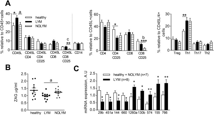
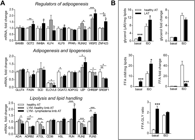
Later stages of secondary lymphedema are associated with the massive deposition of adipose tissue (AT). The factors driving lymphedema-associated AT (LAT) expansion in humans remain rather elusive. We hypothesized that LAT expansion could be based on alterations of metabolic, adipogenic, immune and/or angiogenic qualities of AT. AT samples were acquired from upper limbs of 11 women with unilateral breast cancer-related lymphedema and 11 healthy women without lymphedema. Additional control group of 11 female breast cancer survivors without lymphedema was used to assess systemic effects of lymphedema. AT was analysed for adipocyte size, lipolysis, angiogenesis, secretion of cytokines, immune and stem cell content and mRNA gene expression. Further, adipose precursors were isolated and tested for their proliferative and adipogenic capacity. The effect of undrained LAT- derived fluid on adipogenesis was also examined. Lymphedema did not have apparent systemic effect on metabolism and cytokine levels, but it was linked with higher lymphocyte numbers and altered levels of several miRNAs in blood. LAT showed higher basal lipolysis, (lymph)angiogenic capacity and secretion of inflammatory cytokines when compared to healthy AT. LAT contained more activated CD4+ T lymphocytes than healthy AT. mRNA levels of (lymph)angiogenic markers were deregulated in LAT and correlated with markers of lipolysis. In vitro, adipose cells derived from LAT did not differ in their proliferative, adipogenic, lipogenic and lipolytic potential from cells derived from healthy AT. Nevertheless, exposition of preadipocytes to LAT-derived fluid improved their adipogenic conversion when compared with the effect of serum. This study presents results of first complex analysis of LAT from upper limb of breast cancer survivors. Identified LAT alterations indicate a possible link between (lymph)angiogenesis and lipolysis. In addition, our in vitro results imply that AT expansion in lymphedema could be driven partially by exposition of adipose precursors to undrained LAT-derived fluid.
继发性淋巴水肿的后期阶段与脂肪组织(AT)的大量沉积有关。在人类中,驱动淋巴水肿相关脂肪组织(LAT)扩张的因素仍然相当难以捉摸。我们推测,LAT扩张可能基于AT的代谢、成脂、免疫和/或血管生成特性的改变。从11名患有单侧乳腺癌相关淋巴水肿的女性和11名无淋巴水肿的健康女性的上肢采集AT样本。另外,选取11名无淋巴水肿的女性乳腺癌幸存者作为对照组,以评估淋巴水肿的全身影响。对AT进行分析,检测脂肪细胞大小、脂肪分解、血管生成、细胞因子分泌、免疫和干细胞含量以及mRNA基因表达。此外,分离脂肪前体细胞并检测其增殖和成脂能力。还研究了未引流的LAT衍生液对成脂作用的影响。淋巴水肿对代谢和细胞因子水平没有明显的全身影响,但与血液中较高的淋巴细胞数量和几种miRNA水平的改变有关。与健康的AT相比,LAT表现出更高的基础脂肪分解、(淋巴)血管生成能力和炎性细胞因子分泌。LAT中活化的CD4+ T淋巴细胞比健康的AT更多。(淋巴)血管生成标志物的mRNA水平在LAT中失调,并与脂肪分解标志物相关。在体外,来自LAT的脂肪细胞与来自健康AT的细胞在增殖、成脂、生脂和脂肪分解潜力方面没有差异。然而,与血清的作用相比,前脂肪细胞暴露于LAT衍生液可改善其成脂转化。本研究展示了对乳腺癌幸存者上肢LAT进行首次综合分析的结果。所确定的LAT改变表明(淋巴)血管生成与脂肪分解之间可能存在联系。此外,我们的体外研究结果表明,淋巴水肿中AT的扩张可能部分是由于脂肪前体细胞暴露于未引流的LAT衍生液所致。