Department of Radiology, China-Japan Friendship Hospital, Beijing, China.
Department of Radiology, Baotou Third Hospital, Baotou, the Inner Mongolia Autonomous Region, China.
Korean J Radiol. 2021 Jul;22(7):1124-1131. doi: 10.3348/kjr.2020.0331. Epub 2021 Apr 1.
To evaluate the feasibility, safety, and effectiveness of CT-guided microcoil localization of solitary pulmonary nodules (SPNs) for guiding video-assisted thoracoscopic surgery (VATS).
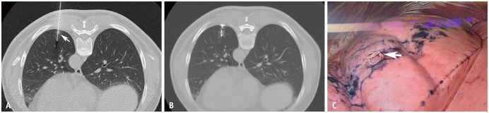
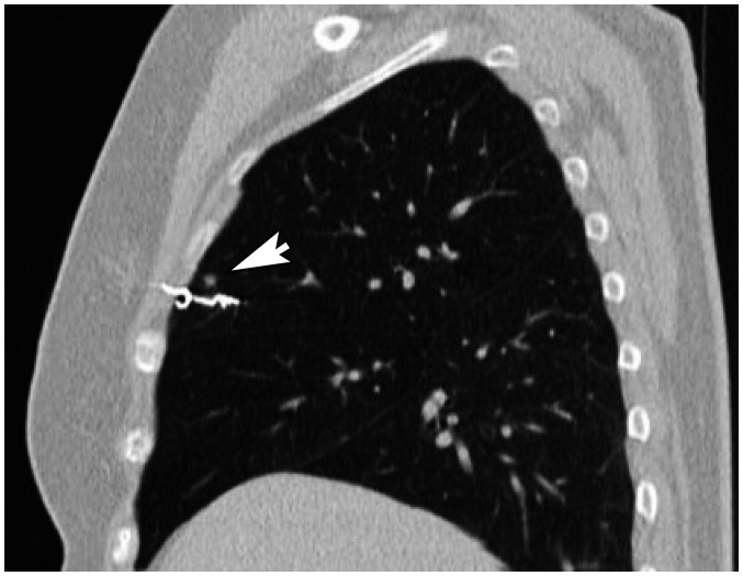
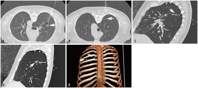
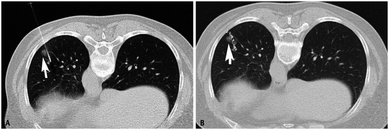
Between June 2016 and October 2019, 454 consecutive patients with 501 SPNs who received CT-guided microcoil localization before VATS in our institution were enrolled. The diameter of the nodules was 0.93 ± 0.49 cm, and the shortest distance from the nodules to the pleura was 1.41 ± 0.95 cm. The distal end of the microcoil was placed less than 1 cm away from the nodule, and the proximal end was placed outside the visceral pleura. VATS was performed under the guidance of implanted microcoils without the aid of intraoperative fluoroscopy.
All 501 nodules were marked with microcoils. The time required for microcoil localization was 12.8 ± 5.2 minutes. Microcoil localization-related complications occurred in 179 cases (39.4%). None of the complications required treatment. A total of 463 nodules were successfully resected under the guidance of implanted microcoils. VATS revealed 38 patients with dislocated microcoils, of which 28 underwent wedge resection (21 cases under the guidance of the bleeding points of pleural puncture, 7 cases through palpation), 5 underwent direct lobectomy, and the remaining 5 underwent a conversion to thoracotomy. In 4 cases, a portion of the microcoil remained in the lung parenchyma.
CT-guided microcoil localization of SPNs is safe and reliable. Marking the nodule and pleura simultaneously with microcoils can effectively guide the resection of SPNs using VATS without the aid of intraoperative fluoroscopy.
评估 CT 引导下微线圈定位孤立性肺结节(SPN)引导电视辅助胸腔镜手术(VATS)的可行性、安全性和有效性。
本研究纳入了 2016 年 6 月至 2019 年 10 月期间在我院接受 CT 引导下微线圈定位后行 VATS 的 454 例连续患者共 501 个 SPN。结节直径为 0.93 ± 0.49cm,结节与胸膜最短距离为 1.41 ± 0.95cm。微线圈的远端放置在距离结节 1cm 以内,近端放置在脏层胸膜外。在植入的微线圈引导下进行 VATS,无需术中透视协助。
所有 501 个结节均被微线圈标记。微线圈定位时间为 12.8 ± 5.2 分钟。179 例(39.4%)发生微线圈定位相关并发症。均无需治疗。在植入的微线圈引导下,共成功切除 463 个结节。VATS 显示 38 例微线圈脱位患者,其中 28 例行楔形切除术(21 例在胸膜穿刺出血点引导下,7 例通过触诊),5 例行直接肺叶切除术,其余 5 例改行开胸术。4 例部分微线圈仍留在肺实质内。
CT 引导下 SPN 微线圈定位安全可靠。微线圈同时标记结节和胸膜,可有效指导 VATS 切除 SPN,无需术中透视。