Scioscia Maria Florencia, Vidal Maritza, Sarli Marcelo, Guelman Rodolfo, Danilowicz Karina, Mana Daniela, Longobardi Vanesa, Zanchetta María Belén
Instituto de Diagnóstico e Investigaciones Metabólicas (IDIM), Universidad del Salvador, ZC 1012 Buenos Aires, Argentina.
Centro de Diagnóstico de Osteoporosis y Enfermedades Reumáticas (CEDOR), San Isidro 15047, Lima, Peru.
J Endocr Soc. 2021 Feb 26;5(5):bvab031. doi: 10.1210/jendso/bvab031. eCollection 2021 May 1.
Pregnancy- and lactation-associated osteoporosis (PLO) is a rare condition characterized by fragility fractures, mostly vertebral, during the third trimester of pregnancy or the early postpartum period.
The aim of this study was to evaluate bone microarchitecture in women with PLO to better understand the pathophysiology of this disease.
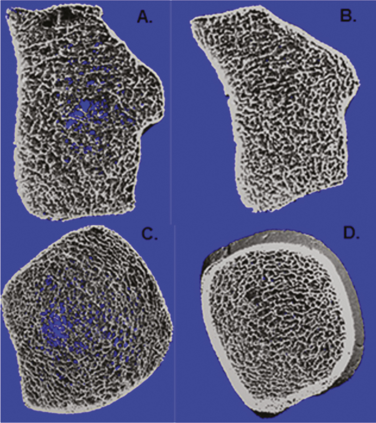
In this retrospective study, we included women with PLO referred to our bone center between November 2007 and July 2012. We assessed bone mineral density (BMD) by dual-energy x-ray absorptiometry, bone turnover markers, and bone microarchitecture by high-resolution peripheral quantitative computed tomography. Results were compared with a control group of healthy lactating women.
Of the 7 primiparous patients with PLO, 6 suffered vertebral fractures and 1 developed a hip fracture during the seventh month of gestation. Fractures occurred within the eighth month of pregnancy and the fourth month post partum; vertebral fractures were multiple in 85.7%. Major or minor risk factors for osteoporosis were present in 86% of our patients. Trabecular density, number, and thickness were 34%, 20% and 22% lower than controls ( < .01, = .01, and = .01, respectively). Cortical parameters were also deteriorated but to a lesser extent.
In comparison with healthy lactating women, patients with PLO presented severe deterioration of bone trabecular and cortical microarchitecture. This significant compromise may explain the occurrence of multiple fractures in these otherwise healthy young women. Further prospective studies are needed to determine whether bone microarchitecture might be able to be restored in the future.
妊娠和哺乳期相关骨质疏松症(PLO)是一种罕见的病症,其特征是在妊娠晚期或产后早期出现脆性骨折,主要是椎体骨折。
本研究的目的是评估PLO女性的骨微结构,以更好地了解该疾病的病理生理学。
在这项回顾性研究中,我们纳入了2007年11月至2012年7月期间转诊至我们骨中心的PLO女性。我们通过双能X线吸收法评估骨密度(BMD)、骨转换标志物,并通过高分辨率外周定量计算机断层扫描评估骨微结构。将结果与健康哺乳期女性对照组进行比较。
7例初产PLO患者中,6例在妊娠第七个月发生椎体骨折,1例发生髋部骨折。骨折发生在妊娠第八个月和产后第四个月内;85.7%的椎体骨折为多发性。86%的患者存在骨质疏松的主要或次要危险因素。小梁密度、数量和厚度分别比对照组低34%、20%和22%(分别为<0.01、=0.01和=0.01)。皮质参数也有所恶化,但程度较轻。
与健康哺乳期女性相比,PLO患者的骨小梁和皮质微结构严重恶化。这种显著的损害可能解释了这些原本健康的年轻女性发生多发性骨折的原因。需要进一步的前瞻性研究来确定未来骨微结构是否能够恢复。