Division of Pediatric Radiology, Hôpital Timone Enfants, Assistance publique - Hôpitaux de Marseille, 264 Rue Sainte Pierre, 13385, Marseille Cedex 05, France.
Division of Pediatric Onco-Hematology, Hôpitaux Universitaires de Genève, Genève, Suisse.
Pediatr Radiol. 2021 Aug;51(9):1714-1723. doi: 10.1007/s00247-021-05037-4. Epub 2021 Apr 20.
Diffusion-weighted imaging (DWI) has been described to correlate with tumoural necrosis in response to preoperative chemotherapy for osteosarcoma.
To assess the accuracy of DWI in evaluating the response to neoadjuvant chemotherapy at the mid-course treatment of long-bone osteosarcoma and in predicting survival.
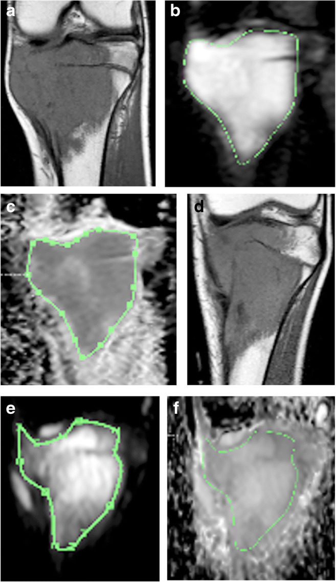
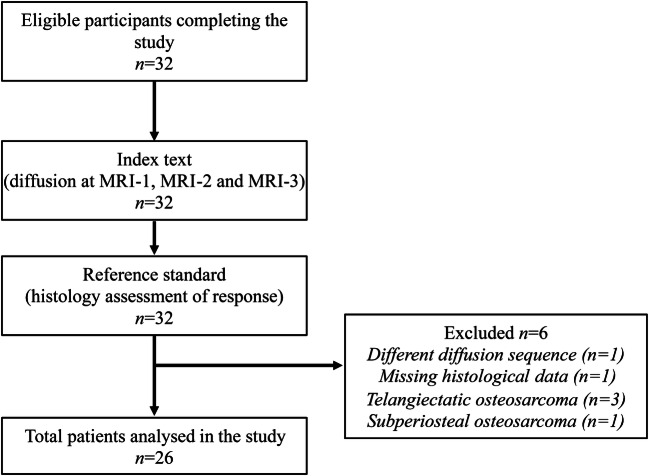
We conducted a prospective single-centre study over a continuous period of 11 years. Consecutive patients younger than 20 years treated with a neoadjuvant regimen for peripheral conventional osteosarcoma were eligible for inclusion. Magnetic resonance imaging (MRI) with DWI was performed at diagnosis, and mid- and end-course chemotherapy with mean apparent diffusion coefficients (ADC) calculated at each time point. A percentage less than or equal to 10% of the viable residual tissue at the histological analysis of the surgical specimen was defined as a good responder to chemotherapy. Survival comparisons were calculated using the Kaplan-Meier method. Uni- and multivariate analyses with ADC change were performed by Cox modelling. This is an expansion and update of our previous work.
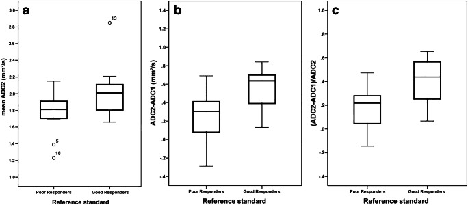
Twenty-six patients between the ages of 4.8 and 19.6 years were included, of whom 14 were good responders. At mid-course chemotherapy, good responders had significantly higher mean ADC values (P=0.046) and a higher increase in ADC (P=0.015) than poor responders. The ADC change from diagnosis to mid-course MRI did not appear to be a prognosticator of survival and did not impact survival rates of both groups.
DWI at mid-course preoperative chemotherapy for osteosarcoma should be considered to evaluate the degree of histological necrosis and to predict survival. The anticipation of a response to neoadjuvant treatment by DWI may have potential implications on preoperative management.
弥散加权成像(DWI)已被描述为与骨肉瘤术前化疗后的肿瘤坏死相关。
评估 DWI 在评估长骨骨肉瘤新辅助化疗中期反应并预测生存方面的准确性。
我们进行了一项为期 11 年的前瞻性单中心研究。符合条件的患者为接受新辅助方案治疗的 20 岁以下外周常规骨肉瘤连续患者。在诊断时进行磁共振成像(MRI)加 DWI,在每个时间点计算平均表观扩散系数(ADC)。在手术标本的组织学分析中,可存活的残留组织百分比小于或等于 10%被定义为对化疗有良好反应者。使用 Kaplan-Meier 方法计算生存比较。通过 Cox 模型进行 ADC 变化的单变量和多变量分析。这是我们以前工作的扩展和更新。
26 名年龄在 4.8 至 19.6 岁之间的患者入选,其中 14 名患者为良好反应者。在中期化疗中,良好反应者的平均 ADC 值明显更高(P=0.046),ADC 增加更高(P=0.015)。从诊断到中期 MRI 的 ADC 变化似乎不是生存的预测因子,也不影响两组的生存率。
骨肉瘤术前新辅助化疗中的 DWI 应考虑用于评估组织学坏死程度并预测生存。DWI 对新辅助治疗的反应预测可能对术前管理具有潜在意义。