Abdulaal Osamah M, McGee Allison, Rainford Louise, O'Driscoll Dearbhail, Galligan Marie, Reid Valerie, MacMahon Peter J
Diagnostic Radiology Technology, College of Applied Medical Sciences, Taibah University, Madina, Saudi Arabia.
Radiography and Diagnostic Imaging, School of Medicine, University College Dublin, Dublin, Ireland.
Insights Imaging. 2021 Apr 20;12(1):54. doi: 10.1186/s13244-021-00992-w.
To investigate the accuracy of Diffusion Weighted Imaging (DWI) using the Readout Segmentation of Long Variable Echo-trains (RESOLVE) sequence in detecting lumbosacral nerve abnormalities.
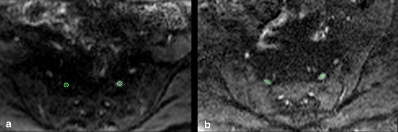
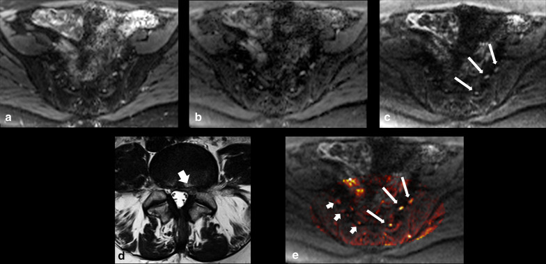
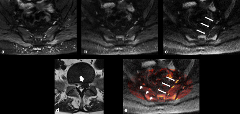
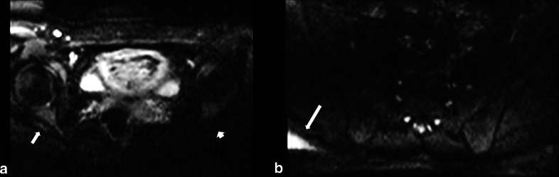
Following institutional ethics committee approval, patients with sciatica-type lower limb radicular symptoms (n = 110) were recruited and prospectively scanned using 3T MRI. Additional participants (n = 17) who underwent neurophysiological testing (EMG/NCV), were also prospectively studied. In addition to routine lumbar spine MRI, a DWI-RESOLVE sequence of the lumbosacral plexus was performed. Two radiologists, blinded to the side of patient symptoms, independently evaluated the MR images. The size and signal intensity changes of the nerves were evaluated using ordinal 4-point Likert-scales. Signal-to-noise ratio (SNR), apparent diffusion coefficient (ADC) and size were measured for affected and normal nerves. Inter-observer agreement was determined with kappa statistics; κ.
In patients who did not undergo EMG/NCV testing (n = 110), the DWI-RESOLVE sequence detected lumbosacral nerve abnormalities that correlated with symptoms in 36.3% (40/110). This is a similar percentage to patients who underwent EMG/NCV testing, which was positive and correlated with symptoms in 41.2% (7/17). Inter-observer agreement for evaluation of lumbosacral nerve abnormalities was excellent and ranged from 0.87 to 0.94. SNR and nerve size measurements demonstrated statistically significant differences for the L5 and S1 nerves (p value < 0.05) for patients who did not undergo EMG/NCV testing.
The DWI-RESOLVE sequence is a promising new method that may permit accurate detection and localization of lumbar nerve abnormalities in patients with sciatica.
探讨采用长可变回波链读出分割(RESOLVE)序列的扩散加权成像(DWI)检测腰骶神经异常的准确性。
经机构伦理委员会批准,招募了有坐骨神经痛型下肢神经根症状的患者(n = 110),并使用3T MRI进行前瞻性扫描。另外17名接受神经生理学测试(肌电图/神经传导速度测定)的参与者也进行了前瞻性研究。除了常规腰椎MRI外,还对腰骶丛进行了DWI-RESOLVE序列扫描。两名对患者症状侧不知情的放射科医生独立评估磁共振图像。使用4级有序李克特量表评估神经的大小和信号强度变化。测量患侧和正常神经的信噪比(SNR)、表观扩散系数(ADC)和大小。观察者间的一致性用kappa统计量(κ)确定。
在未进行肌电图/神经传导速度测定的患者(n = 110)中,DWI-RESOLVE序列检测到与症状相关的腰骶神经异常的比例为36.3%(40/110)。这一比例与接受肌电图/神经传导速度测定的患者相似,后者呈阳性且与症状相关的比例为41.2%(7/17)。观察者间对腰骶神经异常评估的一致性极佳,范围为0.87至0.94。对于未进行肌电图/神经传导速度测定的患者,L5和S1神经的SNR和神经大小测量显示出统计学上的显著差异(p值<0.05)。
DWI-RESOLVE序列是一种有前景的新方法,可能有助于准确检测和定位坐骨神经痛患者的腰神经异常。