Department of Internal Medicine II, Ulm University Medical Center, Albert-Einstein-Allee 23, 89081, Ulm, Germany.
Int J Comput Assist Radiol Surg. 2021 Aug;16(8):1255-1262. doi: 10.1007/s11548-021-02366-5. Epub 2021 Apr 20.
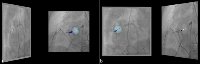
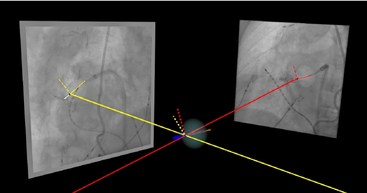
Automatic identification of interventional devices in X-ray (XR) fluoroscopy offers the potential of improved navigation during transcatheter endovascular procedures. This paper presents a prototype implementation of fully automatic 3D reconstruction of a cryo-balloon catheter during pulmonary vein isolation (PVI) procedures by deep learning approaches.
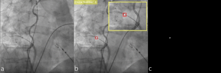
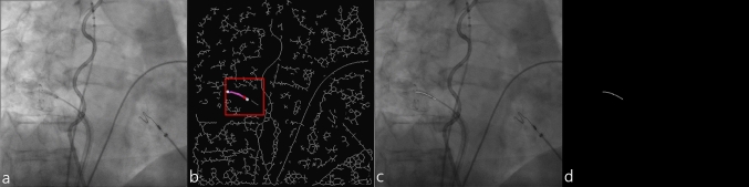
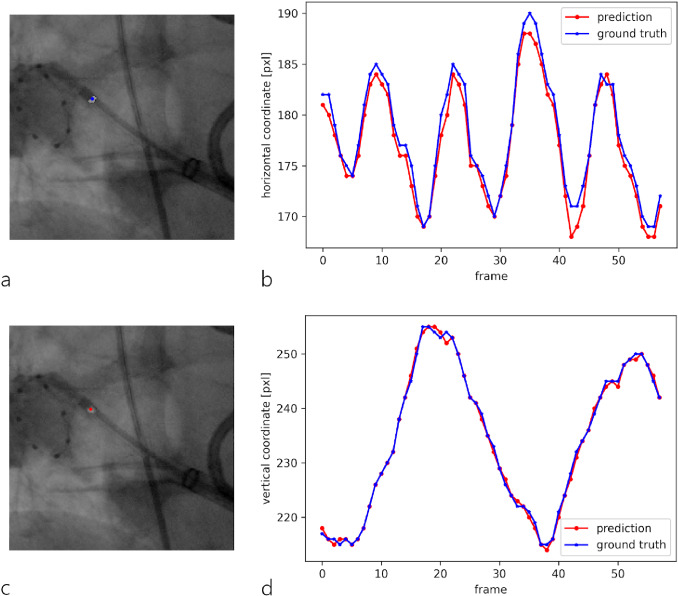
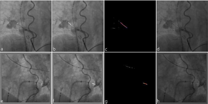
We employ convolutional neural networks (CNN) to automatically identify the cryo-balloon XR marker and catheter shaft in 2D fluoroscopy during PVI. Training data are generated exploiting established semiautomatic techniques, including template-matching and analytical graph building. A first network of U-net architecture uses a single grayscale XR image as input and yields the mask of the XR marker. A second network of the similar architecture is trained using the mask of the XR marker as additional input to the grayscale XR image for the segmentation of the cryo-balloon catheter shaft mask. The structures automatically identified in two 2D images with different angulations are then used to reconstruct the cryo-balloon in 3D.
Automatic identification of the XR marker was successful in 78% of test cases and in 100% for the catheter shaft. Training of the model for prediction of the XR marker mask was successful with 3426 training samples. Incorporation of the XR marker mask as additional input for the model predicting the catheter shaft allowed to achieve good training result with only 805 training samples. The average prediction time per frame was 14.47 ms for the XR marker and 78.22 ms for the catheter shaft. Localization accuracy for the XR marker yielded on average 1.52 pixels or 0.56 mm.
In this paper, we report a novel method for automatic detection and 3D reconstruction of the cryo-balloon catheter shaft and marker from 2D fluoroscopic images. Initial evaluation yields promising results thus indicating the high potential of CNNs as alternatives to the current state-of-the-art solutions.
在 X 射线(XR)透视中自动识别介入设备具有改善经导管血管内程序中导航的潜力。本文通过深度学习方法提出了一种在肺静脉隔离(PVI)程序中自动重建冷冻球囊导管的完全自动 3D 重建的原型实现。
我们使用卷积神经网络(CNN)自动识别 PVI 期间 2D 透视中的冷冻球囊 XR 标记和导管轴。利用已建立的半自动技术(包括模板匹配和分析图构建)生成训练数据。U-net 架构的第一个网络使用单个灰度 XR 图像作为输入,并生成 XR 标记的蒙版。第二个具有类似架构的网络使用 XR 标记的蒙版作为灰度 XR 图像的附加输入进行训练,用于分割冷冻球囊导管轴的蒙版。然后,使用从两个不同角度拍摄的两张自动识别的 2D 图像来重建冷冻球囊的 3D 结构。
XR 标记的自动识别在 78%的测试案例中成功,在 100%的导管轴中成功。用于预测 XR 标记蒙版的模型的训练成功,使用了 3426 个训练样本。将 XR 标记蒙版作为预测导管轴的模型的附加输入进行整合,仅使用 805 个训练样本即可获得良好的训练结果。每个帧的平均预测时间为 XR 标记 14.47 毫秒,导管轴 78.22 毫秒。XR 标记的定位精度平均为 1.52 像素或 0.56 毫米。
在本文中,我们报告了一种从 2D 透视图像自动检测和 3D 重建冷冻球囊导管轴和标记的新方法。初步评估结果令人鼓舞,这表明 CNN 作为当前最先进解决方案的替代方案具有很高的潜力。