Department of Orthopedic Surgery, Tongji Hospital, Tongji Medical College, Huazhong University of Science and Technology, Wuhan 430030, Hubei, China.
Department of Physiology, School of Basic Medicine, Tongji Medical College, Huazhong University of Science and Technology, Wuhan 430030, Hubei, China.
Aging (Albany NY). 2021 Apr 20;13(8):11646-11664. doi: 10.18632/aging.202857.
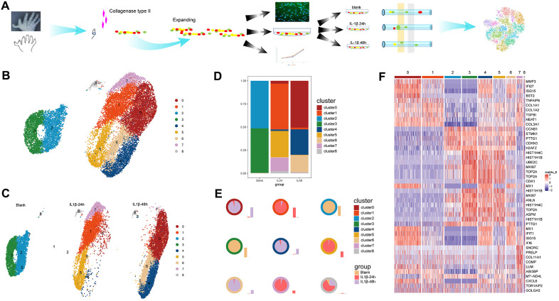
To investigate the heterogeneous responses of expanded chondrocytes, which were cultured in an interleukin (IL)-1β -induced inflammatory environment.
Human articular chondrocytes were expanded, , for 13 days and treated with IL-1β for 0, 24, and 48 h. Cells were collected and subjected to single-cell RNA sequencing. Multiple bioinformatics tools were used to determine the signatures that define chondrocyte physiology.
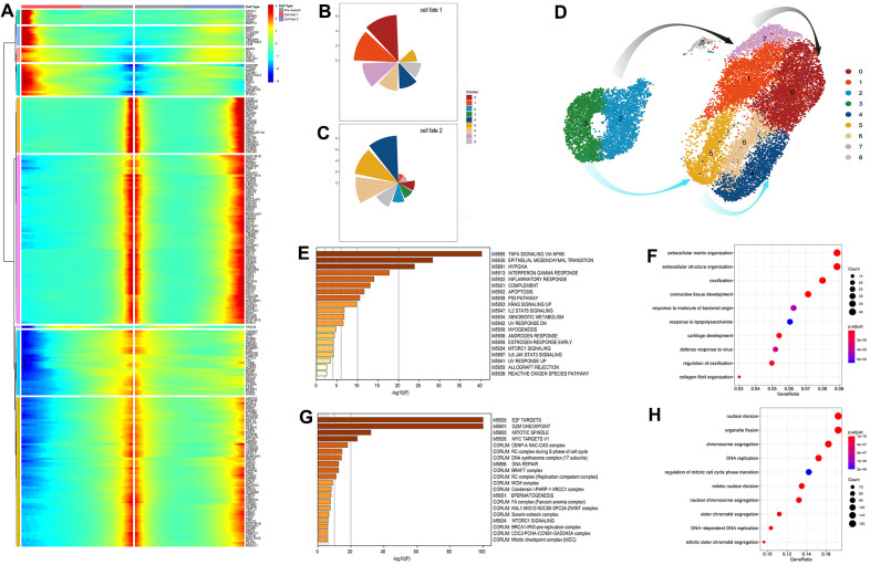
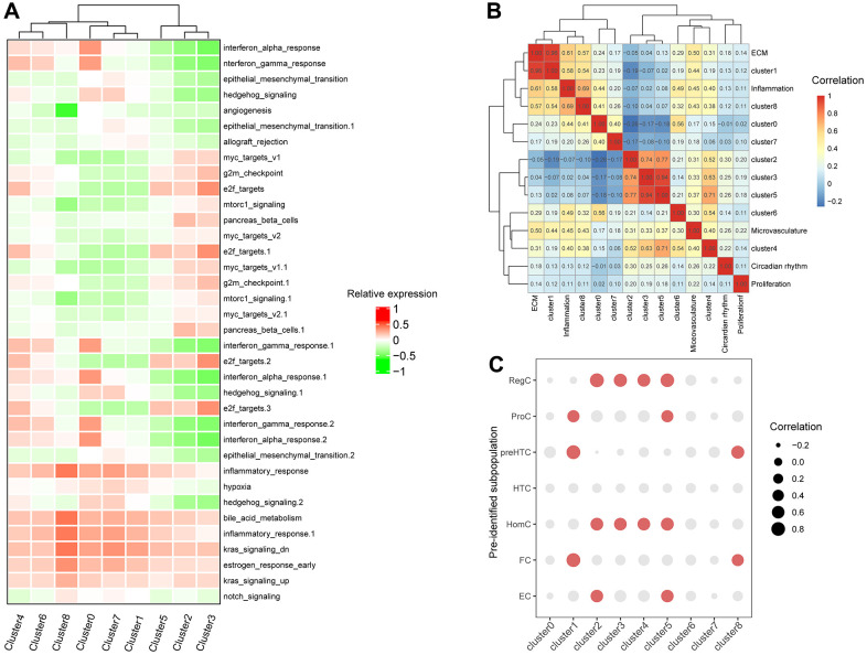
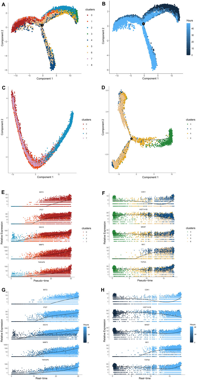
Two major cell clusters with distinct expression patterns were identified at the initial phase and were with heterogeneous variation that coincides with inflammation progress. They transformed into two terminal cell clusters one of which exhibited OA-phenotype and proinflammatory characteristics through two paths, "response-to-inflammation" and "atypical response-to-inflammation", respectively. The involved cell clusters exhibited intrinsic relationship with cell types within native cartilage from OA patients. Genes controlling cell transformation to OA-phenotype were relating to the tumor necrosis factor (TNF) signaling pathway via NFKB, up-regulated KRAS signaling and the IL2/STAT5 signaling pathway and pathways relating to apoptosis and reactive oxygen species.
The expanded chondrocytes under IL-1β-induced inflammatory progression behave heterogeneously. One of the initial cell clusters could transform into a proinflammatory subpopulation through a termed response-to-inflammation path, which may serve as the core target to alleviate OA progression.
研究在白细胞介素(IL)-1β诱导的炎症环境中培养的扩增软骨细胞的异质反应。
扩增人关节软骨细胞,培养 13 天,用 IL-1β处理 0、24 和 48 小时。收集细胞并进行单细胞 RNA 测序。使用多种生物信息学工具来确定定义软骨细胞生理学的特征。
在初始阶段鉴定出具有不同表达模式的两个主要细胞簇,并且存在与炎症进展一致的异质变化。它们通过两条途径转化为两个终末细胞簇,其中一个通过“炎症反应”和“非典型炎症反应”分别表现出 OA 表型和促炎特征。所涉及的细胞簇与 OA 患者原生软骨内的细胞类型具有内在关系。控制细胞向 OA 表型转化的基因通过 NFKB 与肿瘤坏死因子(TNF)信号通路相关,上调 KRAS 信号和 IL2/STAT5 信号通路以及与细胞凋亡和活性氧相关的通路。
在 IL-1β诱导的炎症进展下扩增的软骨细胞表现出异质性。初始细胞簇之一可通过所谓的炎症反应途径转化为促炎亚群,这可能是缓解 OA 进展的核心靶点。