Fadzli Farhana, Rahmat Kartini, Ramli Marlina Tanty, Rozalli Faizatul Izza, Hooi Teoh Kean, Fadzli Ahmad Nazran, Hoong See Mee, Ramli Norlisah Mohd, Taib Nur Aishah Mohd
Department of Biomedical Imaging, University Malaya Research Imaging Centre, University of Malaya, Kuala Lumpur.
Radiology Department, Faculty of Medicine, University Teknologi MARA, Sungai Buloh Campus, Selangor.
Medicine (Baltimore). 2021 Apr 23;100(16):e25297. doi: 10.1097/MD.0000000000025297.
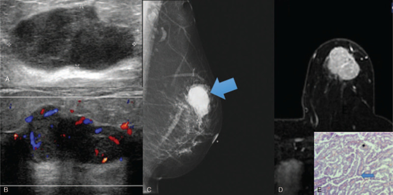
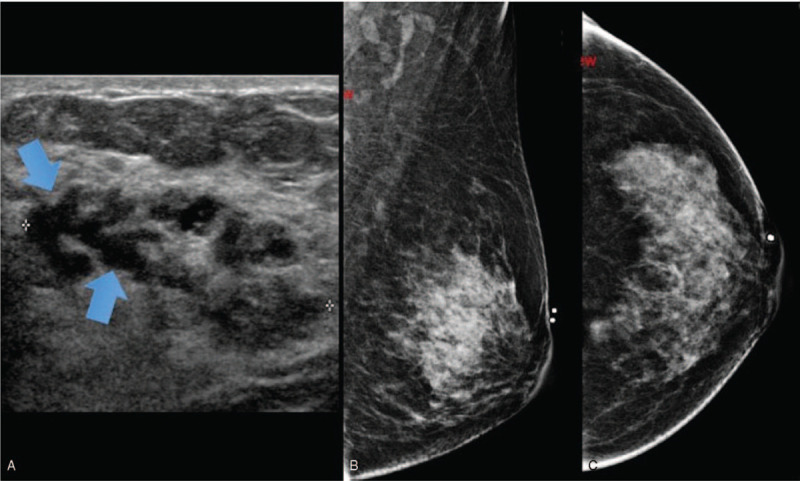
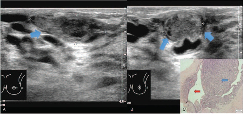
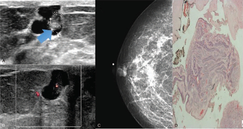
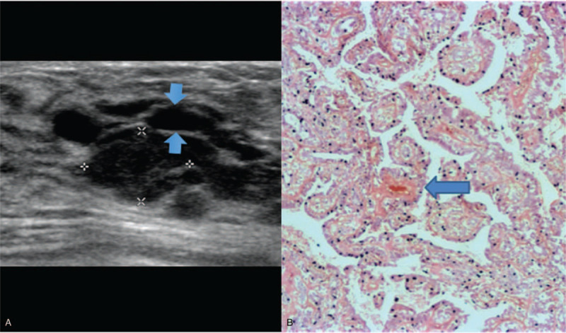
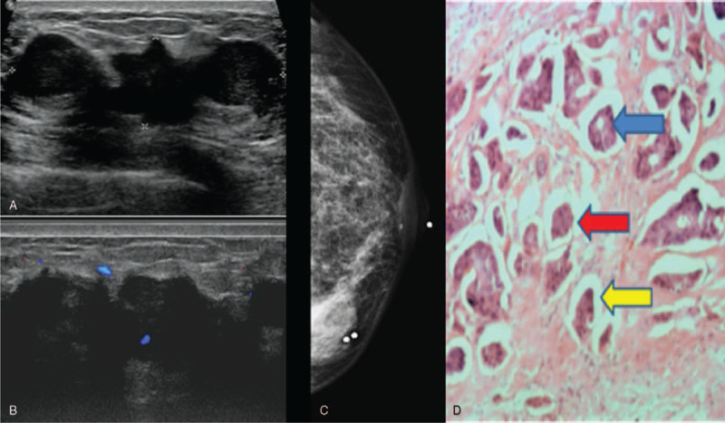
To evaluate the clinical and imaging findings of papillary breast neoplasm and review the pathologic correlation at a tertiary center.Retrospective study of patients diagnosed with benign and malignant papillary lesions between 2008 to 2018. 147 patients were identified with histology diagnosis of papillary lesions. The clinical, imaging, and pathological characteristics were reviewed.Patient cohort included 147 women diagnosed with papillary lesions (mean age at diagnosis 53.8 years) and were divided into 3 histology groups (benign, atypical, and malignant). Common clinical presentations were breast lump (n = 60) and nipple discharge (n = 29), 48 patients were asymptomatic.Only 37 were detected as a mass lesion on mammogram. The presence of mass lesion on mammogram was the most common feature in all 3 papillary lesion groups, and with the presence of asymmetric density, were the 2 mammographic features significantly associated (P < .05) with malignancy.All lesions were detected on ultrasound. The most common sonographic features for all 3 groups were the presence of a mass and irregular shape. Among all the sonographic features assessed, larger size, presence of vascularity and absence of dilated ducts were significantly associated (P < .05) with malignancy.Feature pattern recognition of the variety of benign, atypical and malignant papillary neoplasm on ultrasound and mammogram, with emphasis on size, presence of vascularity and dilated ducts on ultrasound and presence of mass, and architectural distortion on mammogram, is important in the assessment of patients with suspected ductal lesions to facilitate optimal treatment and surgical care.
评估乳腺乳头状肿瘤的临床和影像学表现,并在三级医疗中心回顾其病理相关性。对2008年至2018年间诊断为良性和恶性乳头状病变的患者进行回顾性研究。确定了147例经组织学诊断为乳头状病变的患者。回顾其临床、影像学和病理特征。患者队列包括147例诊断为乳头状病变的女性(诊断时平均年龄53.8岁),分为3个组织学组(良性、非典型和恶性)。常见临床表现为乳腺肿块(n = 60)和乳头溢液(n = 29),48例患者无症状。乳腺钼靶检查仅37例被检测为肿块病变。乳腺钼靶检查发现肿块病变是所有3个乳头状病变组中最常见的特征,且与密度不对称一起,是与恶性肿瘤显著相关(P < 0.05)的2个乳腺钼靶特征。所有病变在超声检查中均被检测到。所有3组最常见的超声特征是存在肿块和形状不规则。在评估的所有超声特征中,较大尺寸、存在血管和无扩张导管与恶性肿瘤显著相关(P < 0.05)。在超声和乳腺钼靶上识别各种良性、非典型和恶性乳头状肿瘤的特征模式,重点是超声上的大小、血管存在和扩张导管以及乳腺钼靶上的肿块存在和结构扭曲,对于评估疑似导管病变的患者以促进最佳治疗和手术护理很重要。