Wang Yingjiao, Li Yuechong, Qu Yang, Zhou Yidong, Sun Qiang, Shen Songjie
Department of Breast Surgery, Peking Union Medical College Hospital, Peking Union Medical College, Chinese Academy of Medical Sciences, 1 Shuaifuyuan, Dongcheng District, Beijing, 100730, China.
Discov Oncol. 2024 Dec 23;15(1):831. doi: 10.1007/s12672-024-01681-y.
Intraductal papillary neoplasms (IPNs) often have a similar clinical and imaging presentation, making them difficult to diagnose. We designed this study to refine and compare intraductal papillary neoplasms' clinical and imaging characteristics.
This study included a total of 154 patients with a postoperative diagnosis of IPNs and collected their clinical, imaging, and pathological data. We compared the clinical and imaging characteristics of benign, atypical hyperplasia, and malignant lesions. We also compared the diagnostic efficacy of ultrasound and mammography.
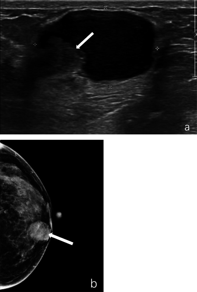
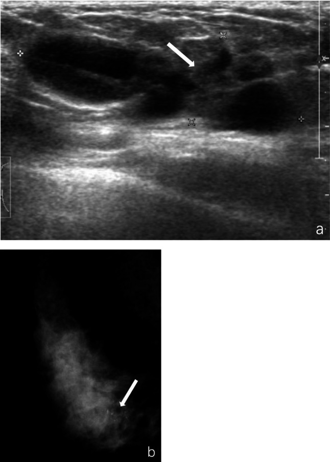
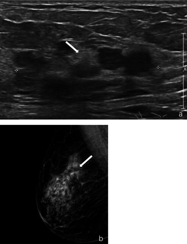
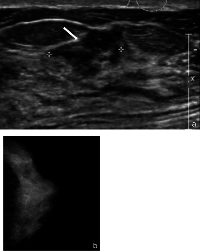
The mean age of malignant patients was 57 years old, which was significantly higher than that in the other groups (48 years in the benign group and 47 years in the atypical hyperplasia group). The proportion of patients with malignant lesions clinically presenting as palpable masses (31.3%) was significantly higher than that of benign lesions (8.6%) (P < 0.05). The proportion of malignant lesions presenting in the periphery (≥ 3 cm from the nipple) was 40.6% compared to 22.4% for benign (P < 0.05). In ultrasonography, characteristics that showed statistically significant differences between benign and malignant lesions were the shape of the mass and calcification (P < 0.05). On mammography, differences were found in mass shape, calcification, and density of masses and glands (P < 0.05).
Clinical features such as age, symptoms, lesion location, and imaging characteristics such as shape, calcification, mass, and density may help to differentiate the classifications of IPNs.
This study was registered at ClinicalTrials.gov on 12/06/2020 (identifier: NCT04429269).
导管内乳头状肿瘤(IPNs)通常具有相似的临床和影像学表现,难以诊断。我们设计了本研究以细化并比较导管内乳头状肿瘤的临床和影像学特征。
本研究共纳入154例术后诊断为IPNs的患者,收集其临床、影像学和病理数据。我们比较了良性、非典型增生和恶性病变的临床和影像学特征。我们还比较了超声和乳腺X线摄影的诊断效能。
恶性患者的平均年龄为57岁,显著高于其他组(良性组为48岁,非典型增生组为47岁)。临床上表现为可触及肿块的恶性病变患者比例(31.3%)显著高于良性病变患者(8.6%)(P<0.05)。恶性病变出现在外周(距乳头≥3 cm)的比例为40.6%,而良性病变为22.4%(P<0.05)。在超声检查中,良性和恶性病变之间显示出统计学显著差异的特征是肿块形状和钙化(P<0.05)。在乳腺X线摄影中,在肿块形状、钙化以及肿块和腺体密度方面发现了差异(P<0.05)。
年龄、症状、病变位置等临床特征以及形状、钙化、肿块和密度等影像学特征可能有助于区分IPNs的分类。
本研究于2020年6月12日在ClinicalTrials.gov注册(标识符:NCT04429269)。